
|
|
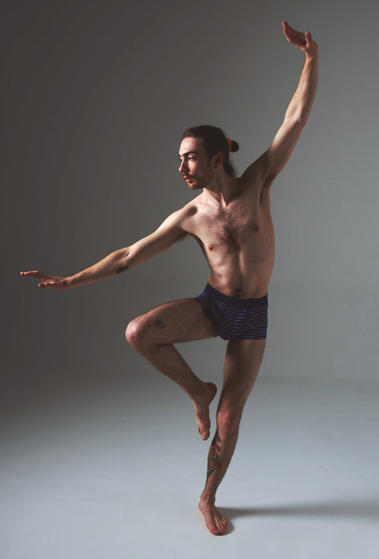
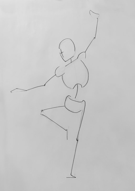
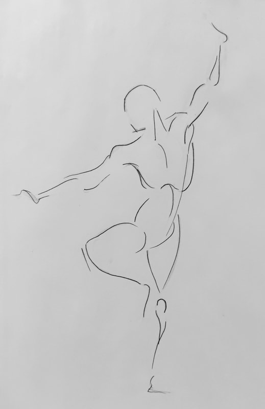
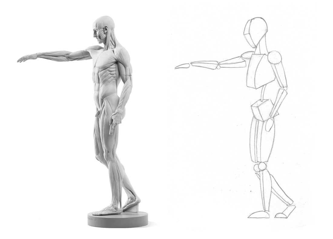
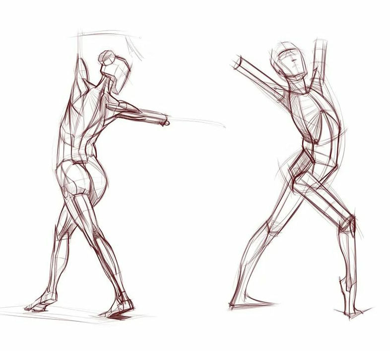
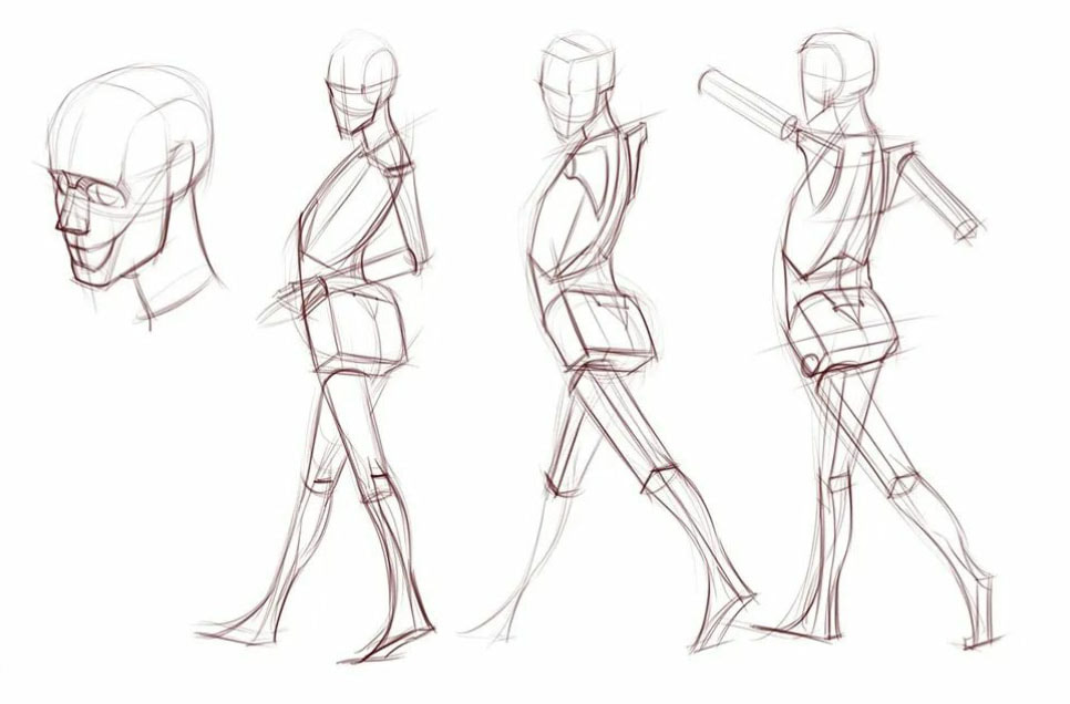
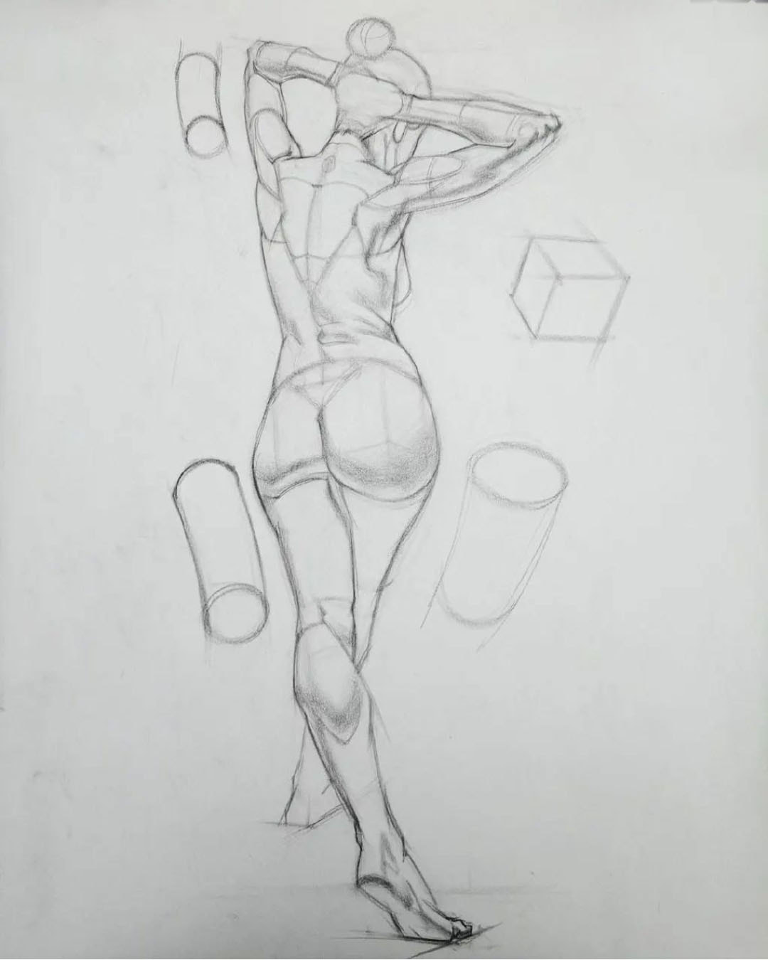
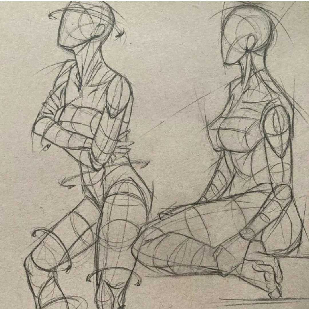
Gesture
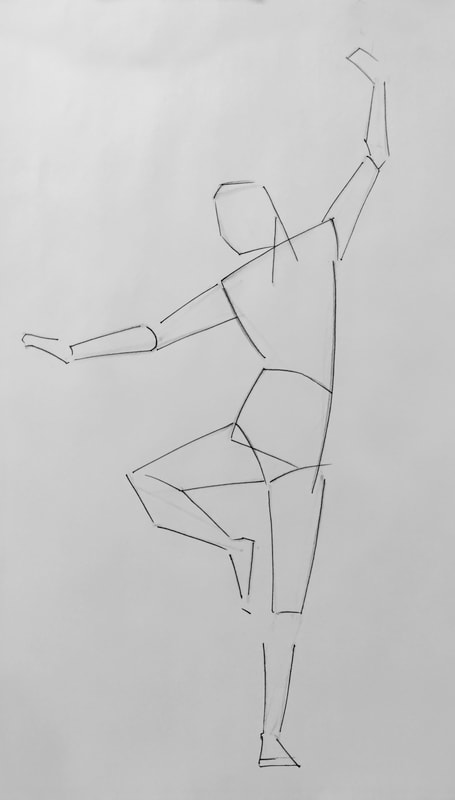
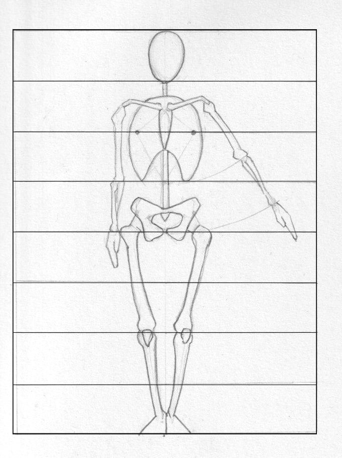
General proportions of the body
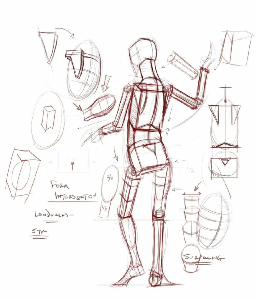
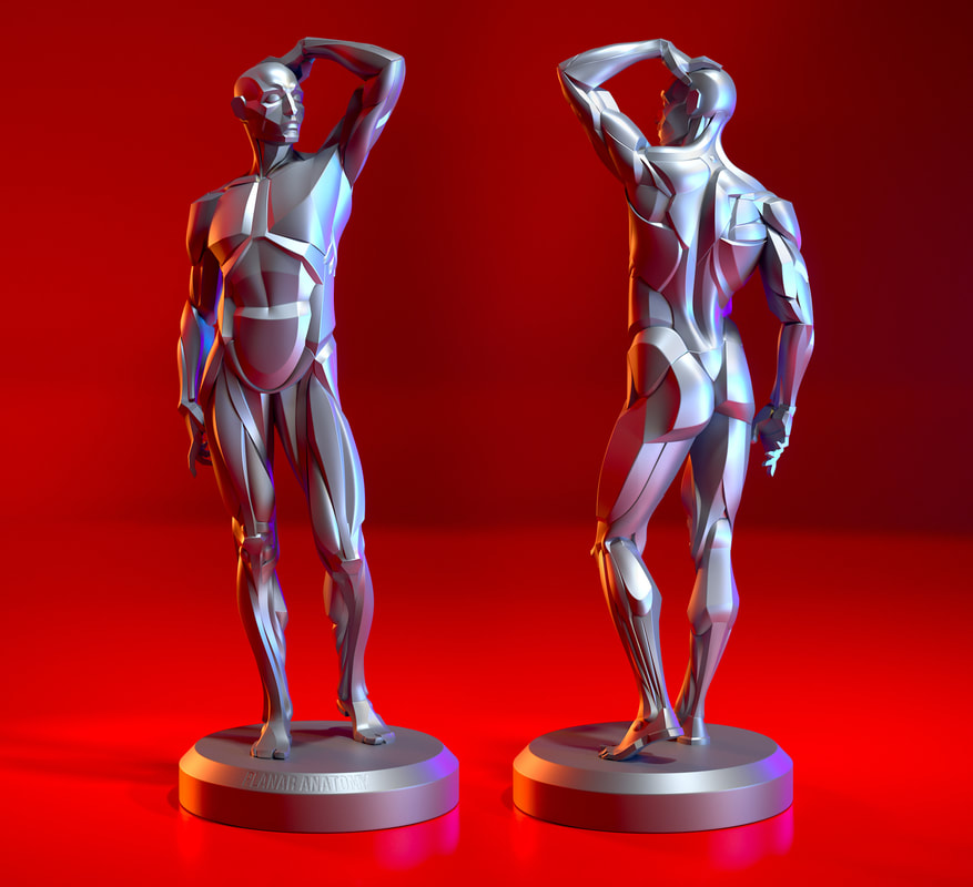
Structure


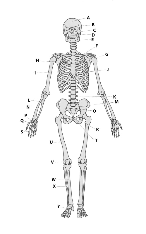
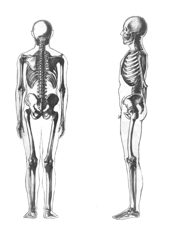
Overview of the bones of the body
|
A - Frontal Bone B - Parietal Bone C - Zygomatic bone D - Maxilla E - Mandible F - Clavicle G - Scapula H - Sternum I - Humerus J - Rib cage K - Spine L - Ulna M - Sacrum N - Radius O - Illium P - Carpals Q - Metacarpals R - Ischium S - Phalanges T - Pubic Bone U - Femur V - Patella W - Tibia X - Fibula Y - Tarsals |
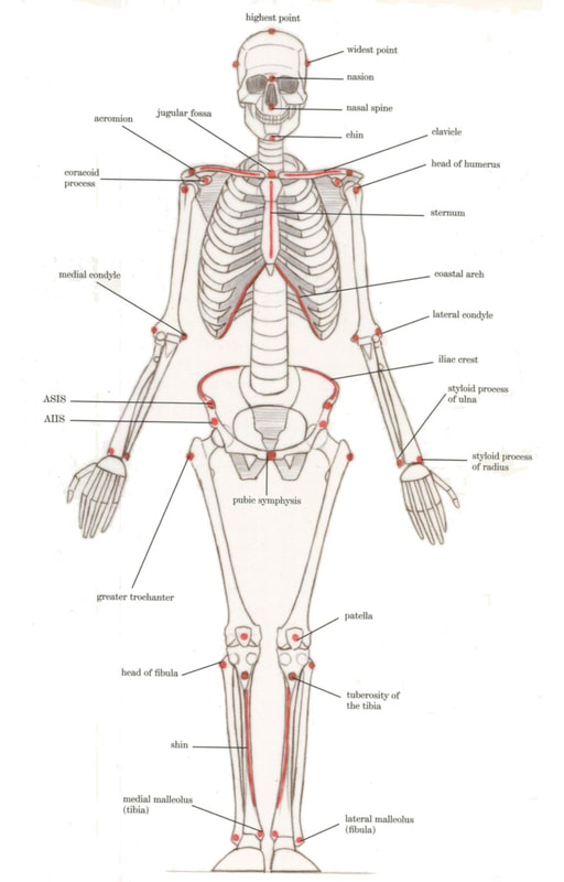
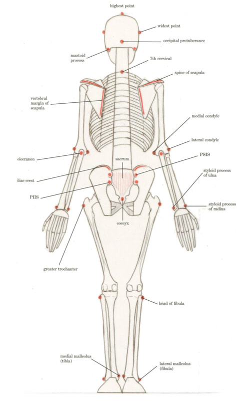
Landmarks of the Skeleton
Below are images from Robert Osti's book " Basic Human Anatomy."
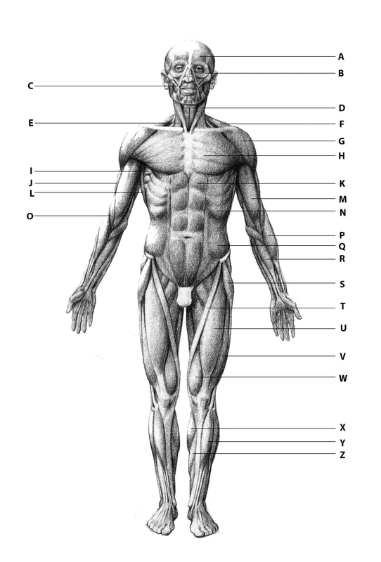
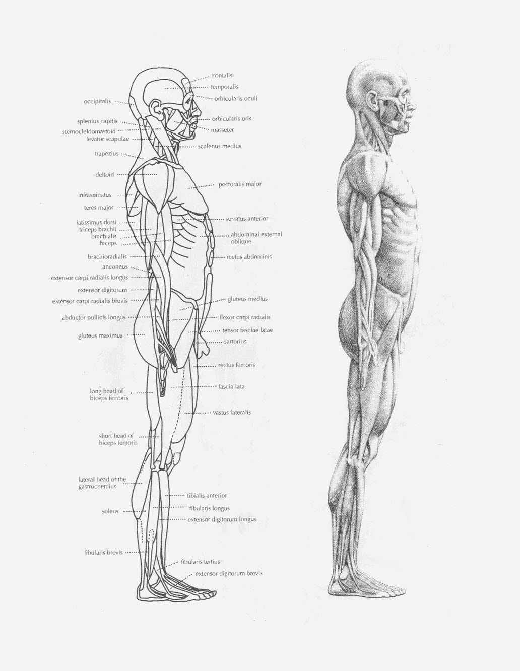
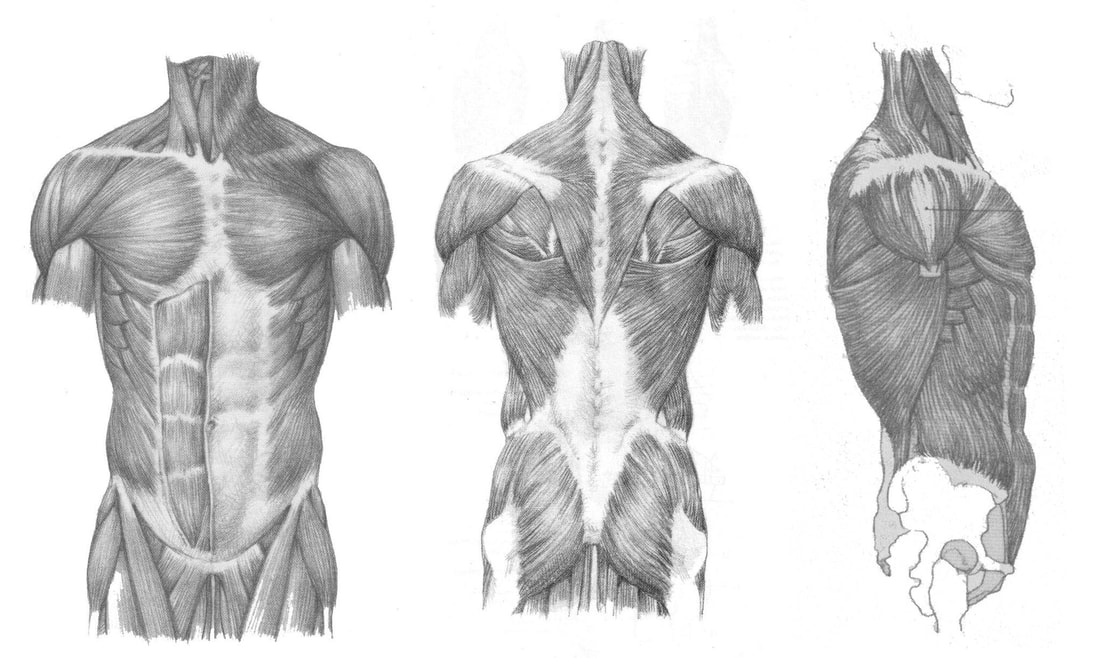
Overview of the muscles of the body
|
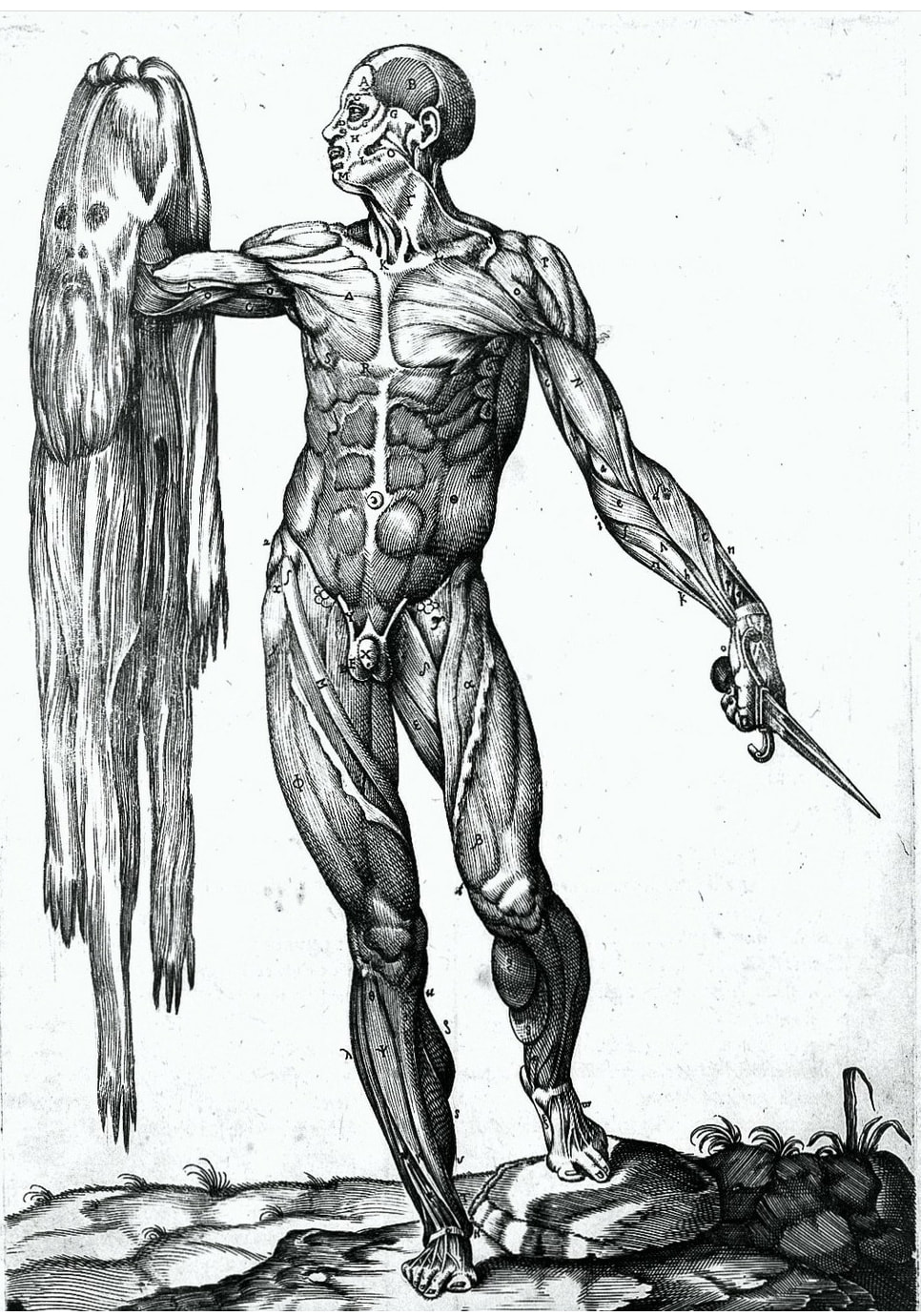
A - Frontalis B - Orbicularis Oculi C - Orbiuclaris Oris D - Sternocleidomastoid E - Sternocleidomastoid F - Trapezius G - Deltoid H - Pectoralis Major I - Serratus Anterior J - Triceps Brachii K - Rectus Abdominus L - Latissimus Dorsi M - Biceps Brachii N - Abdominal External Oblique O - Brachialis P - Brachioradialis Q - Abdominal External Oblique R - Flexor Carpi Radialis S - Tensor Fascia Lata T - Sartorius U - Rectus Femoris V - Vastus Lateralis W - Vastus Medialis X - Gasrtocnemius Y - Tibialis Anterior Z - Soleus |
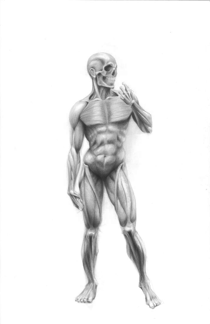
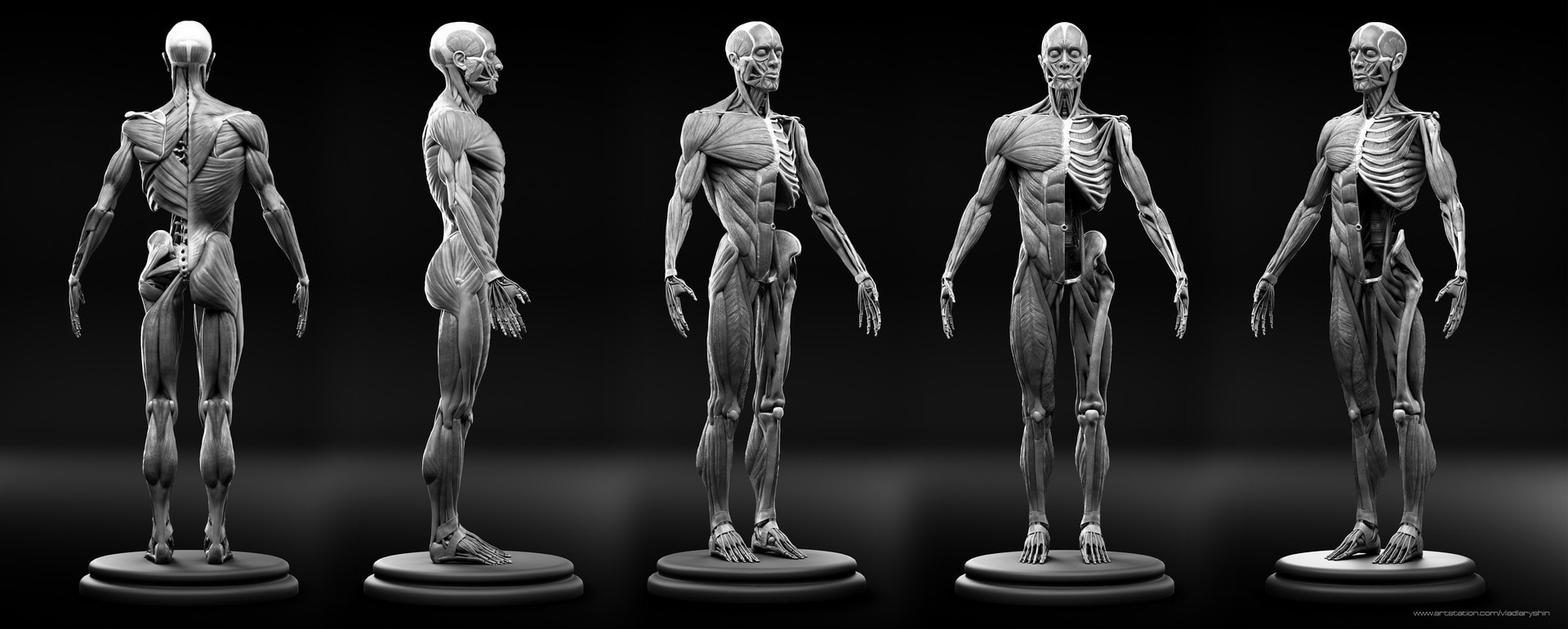
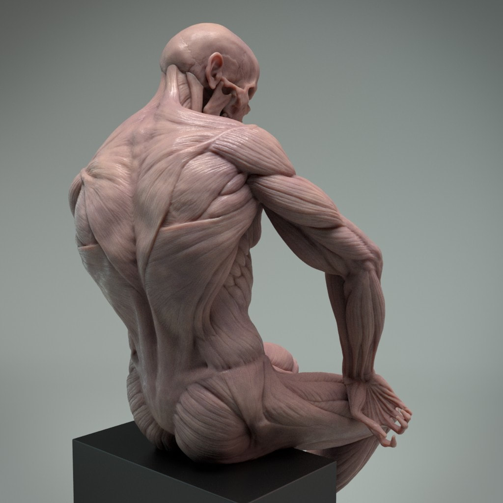
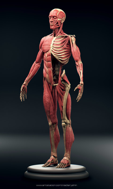
Below is full body ecorche by Vladislav Laryushi
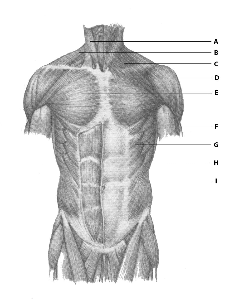
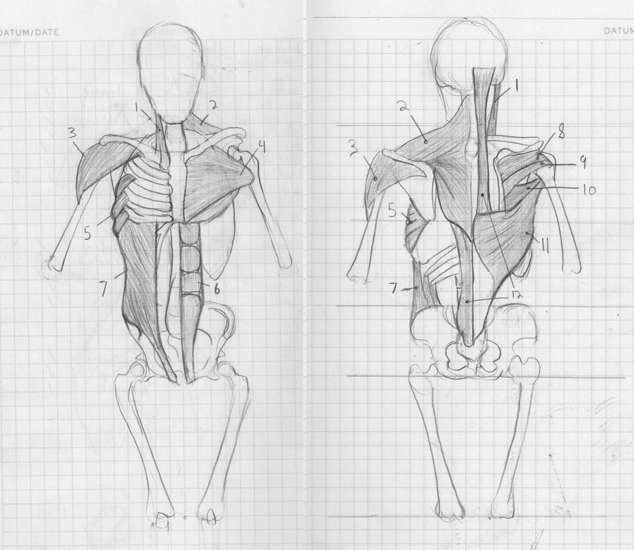
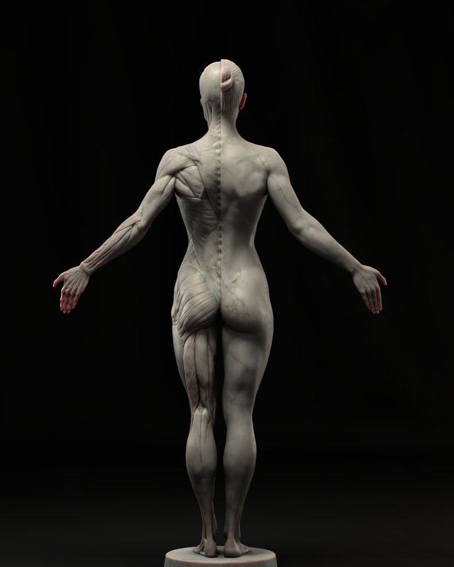
Torso
J - Trapezius - tilting the scapula, helping to life the arm
K - Deltoid - rotating the arm L - Infraspinatus - rotating the arm M - Teres Minor - lowering and rotating the arm N - Teres Major - lowering and rotating the arm O - Latissimus Dorsi - lowering and rotating the arm P - Abdominal External Oblique - bending and rotating the torso Q - Gluteus Medius - abducting the thigh R - Gluteus Maximus - extending the thigh |
A - Sternocleidomastoid - tilting and rotating the head B - Trapezius - tilting the scapula, helping to life the arm C- Platysma - covers the muscles of the neck D - Deltoid - rotating the arm E - Pectoralis Major - lowering and adducting the arm F - Serratus Anterior - tilting the scapula, helping to lift the arm G - Abdominal External Oblique - bending and rotating the torso H - Rectus Sheath - covers the muscles of the abdomen I - Rectus Abdominus - flexing the torso 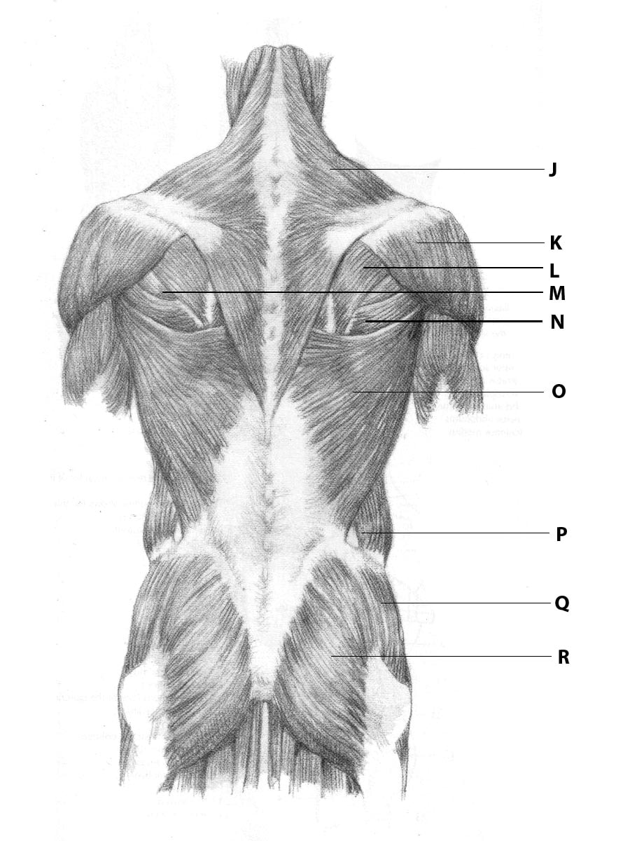
|
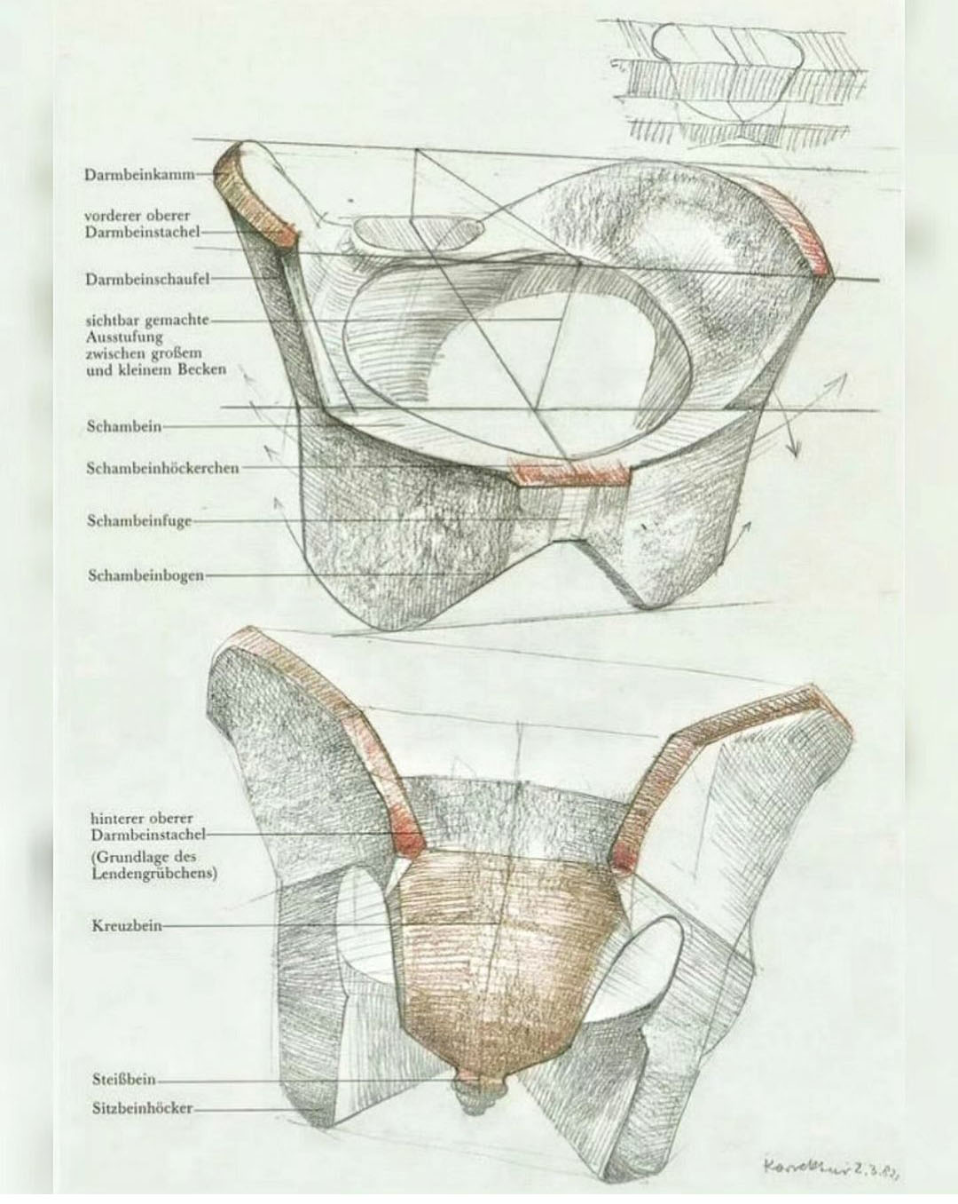
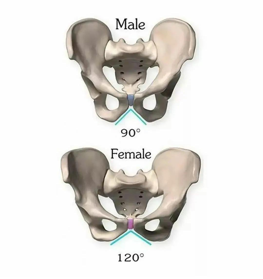
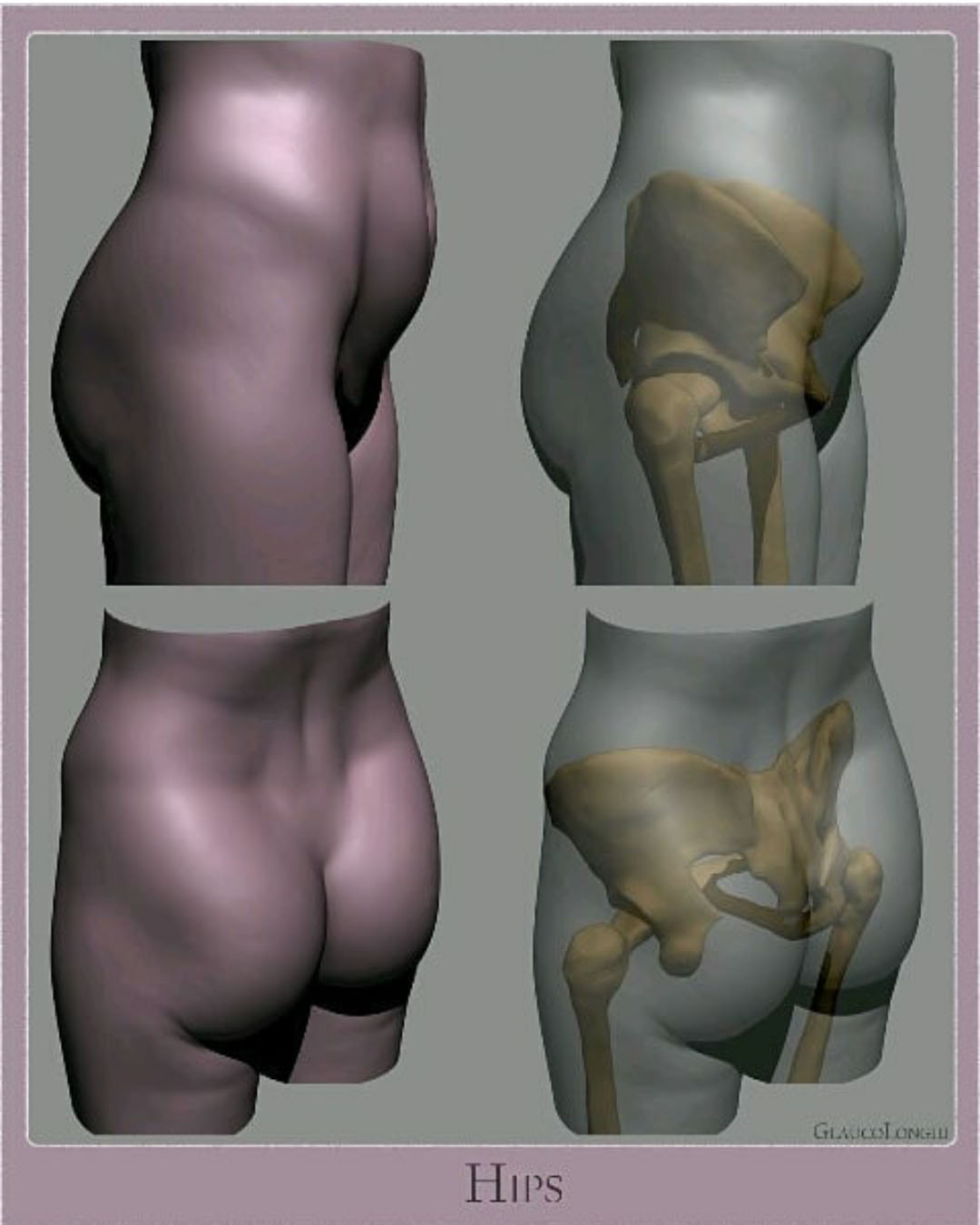
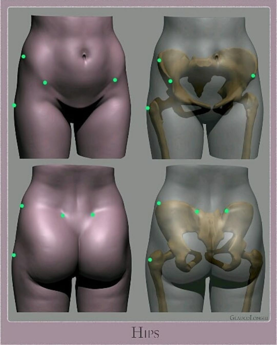
Pelvis
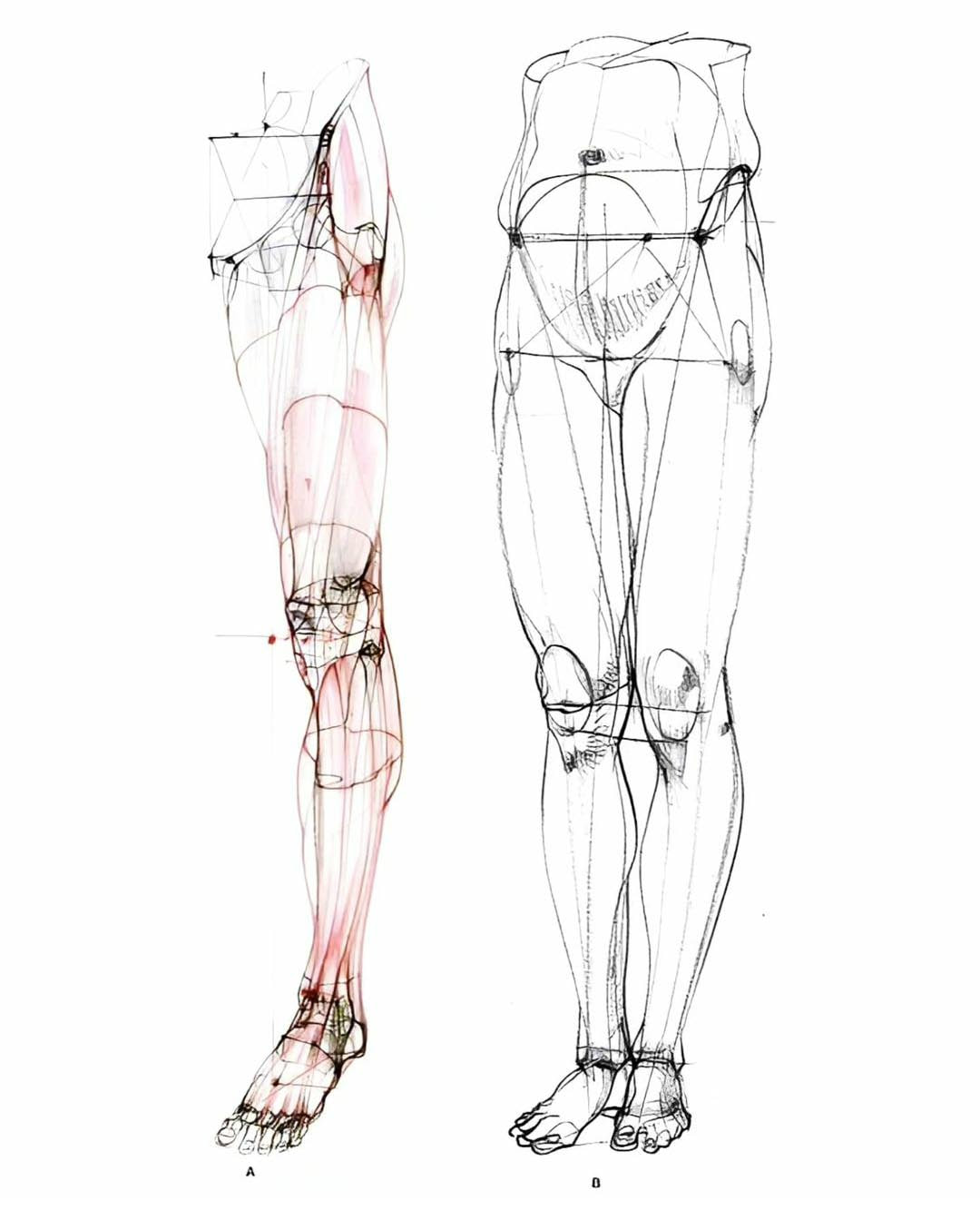
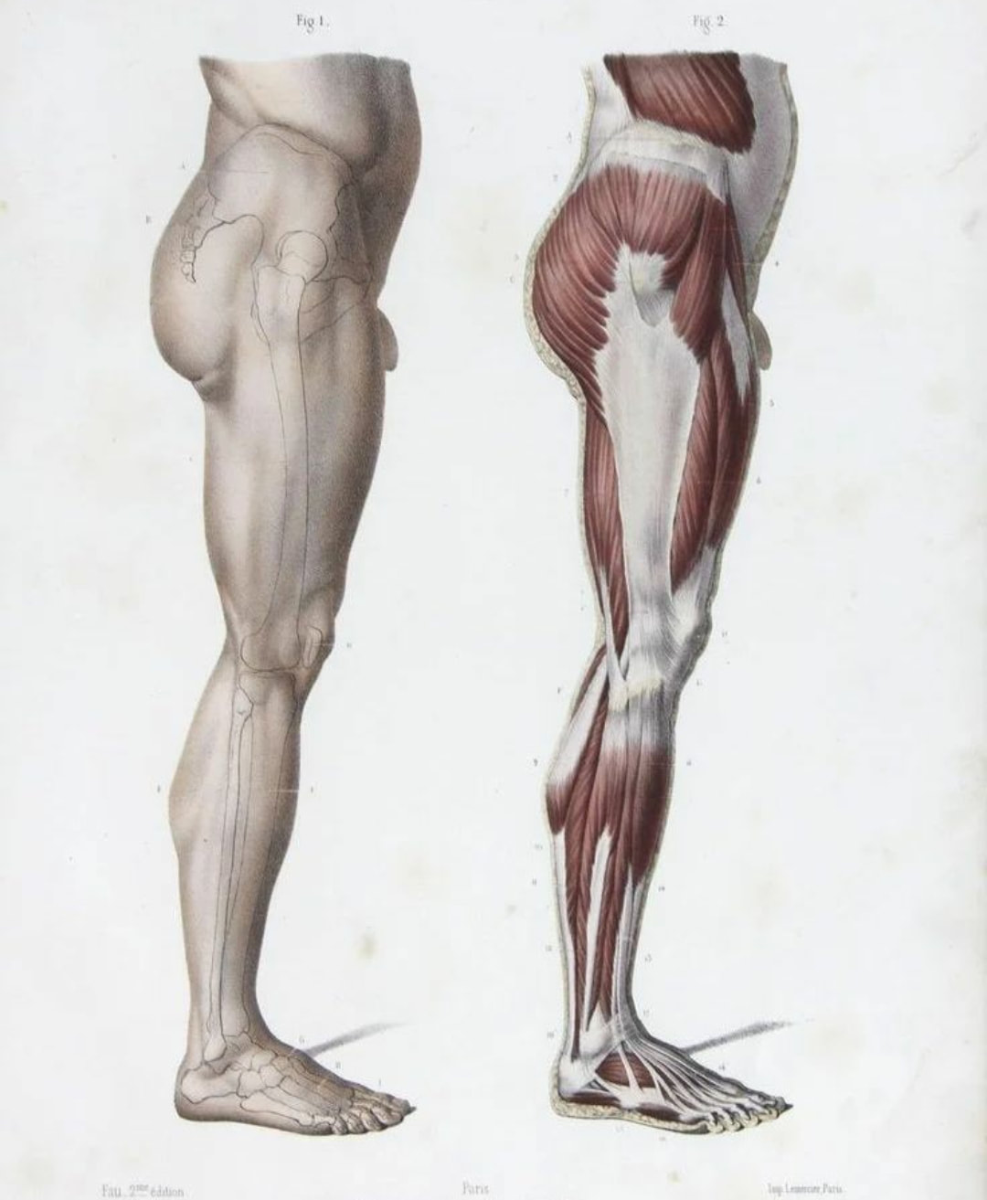
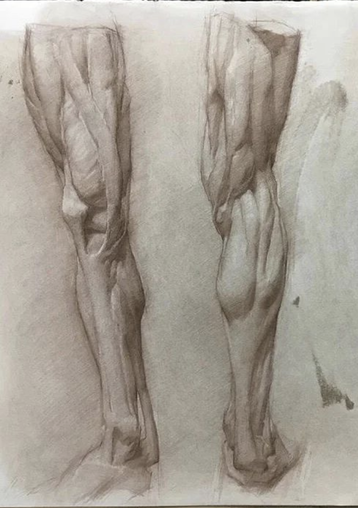
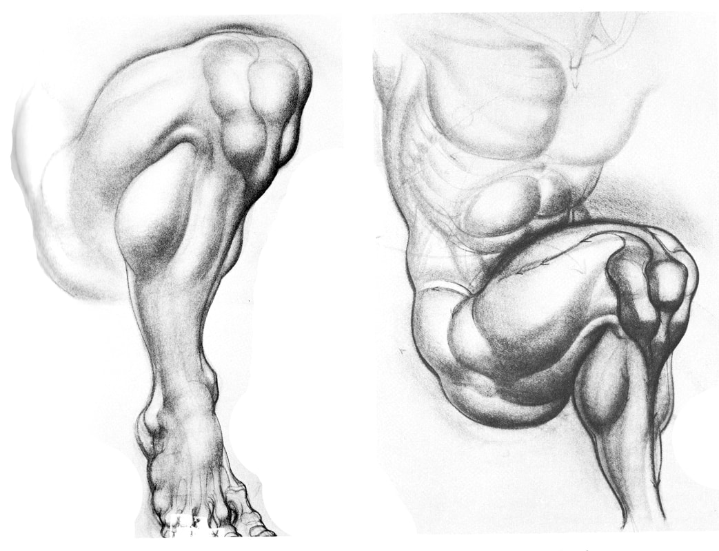
Legs
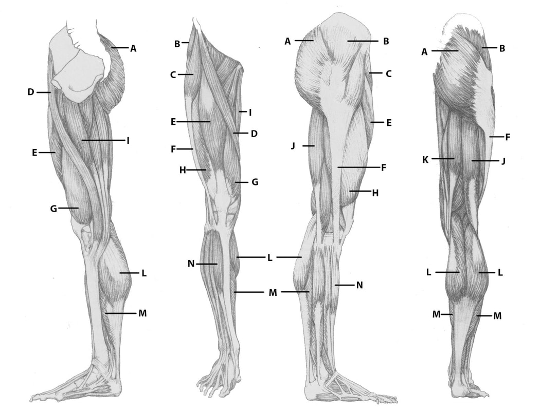
E, G, & H - Quadriceps- Begin at the Illium and femur and all connect at the tendon around the patella, and then insert at the tibia
J & K - Hamstrings - Begin at the pelvis and insert the tibia (semitendinosus) and fibula (bicep femoris) |
A - Gluteus Maximus - ins. pelvis, femur - extending the thigh,
B - Gluteus Medialis - ins. pelvis, femur - abducting the thigh C - Tensor Fascia Latae - ins. pelvis, tibia - flexing the thigh D - Sartorius - ins. pelvis, tibia - flexing and rotating the thigh and leg E - Rectus Femoris - flexing the thigh, extending the leg (quadricep) F - Fascia Lata - Helps to steady and stabilize knee and hip joints G - Vastus Medialis - flexing the thigh, extending the leg (quadricep) H - Vastus Lateralis - flexing the thigh, extending the leg (quadricep) I - Gracilis - ins. pelvis, tibia - adducting the thigh J - Biceps Femoris - flexing the leg (hamstring) K - Semitendinosus - flexing the leg (hamstring) L - Gastrocnemius - ins. femur, heel - extending the foot M - Soleus - ins. tibia, fibula - extending the foot N - Anterior Tibialis - ins. Tibia, 1st metatarsal - flexing the foot |
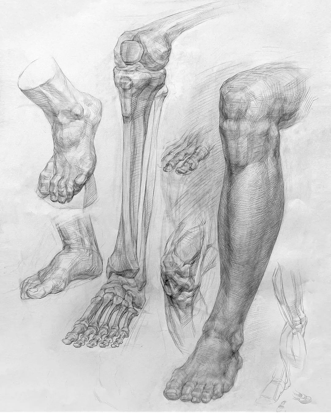
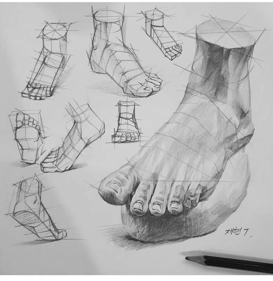
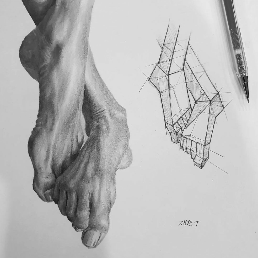
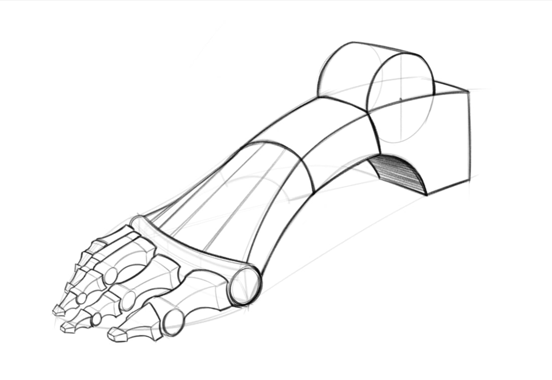
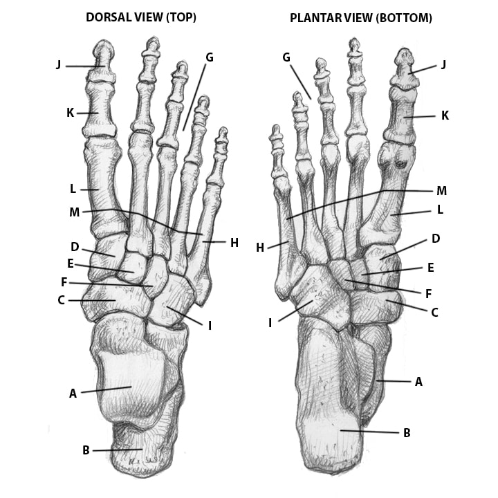
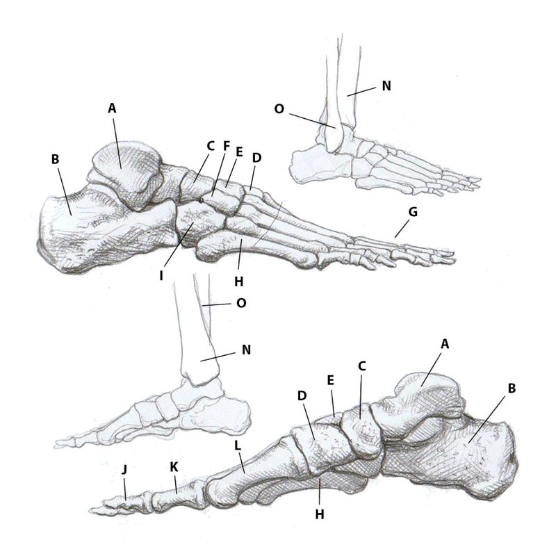
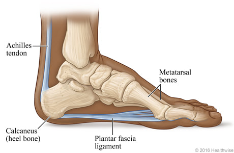
fEET
|
A - Astragalus / Talus
B - Calcaneus (Heel) C - Scaphoid D - 1st Cuneiform E - 2nd Cuneiform F - 3rd Cuneiform G - Phalanges H - 1st Metatarsal |
I - Cuboid
J - Distal Phalanx of the big toe K - Proximal Phalanx of the big toe L - 1st Metatarsal M - Metatarsal Bones N - Tibia O - Fibula |
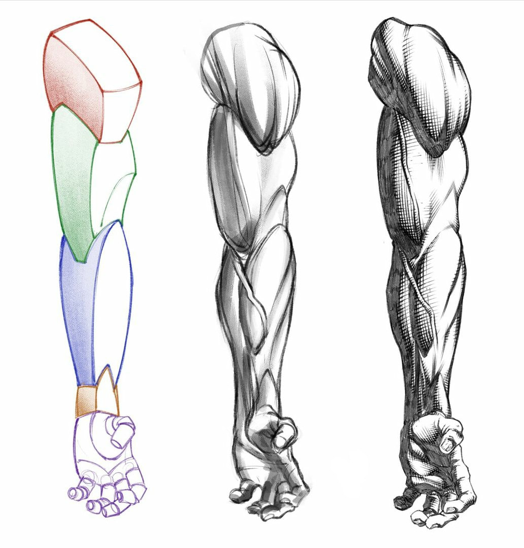
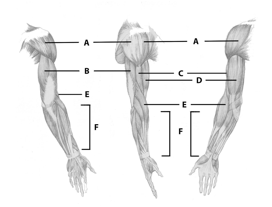
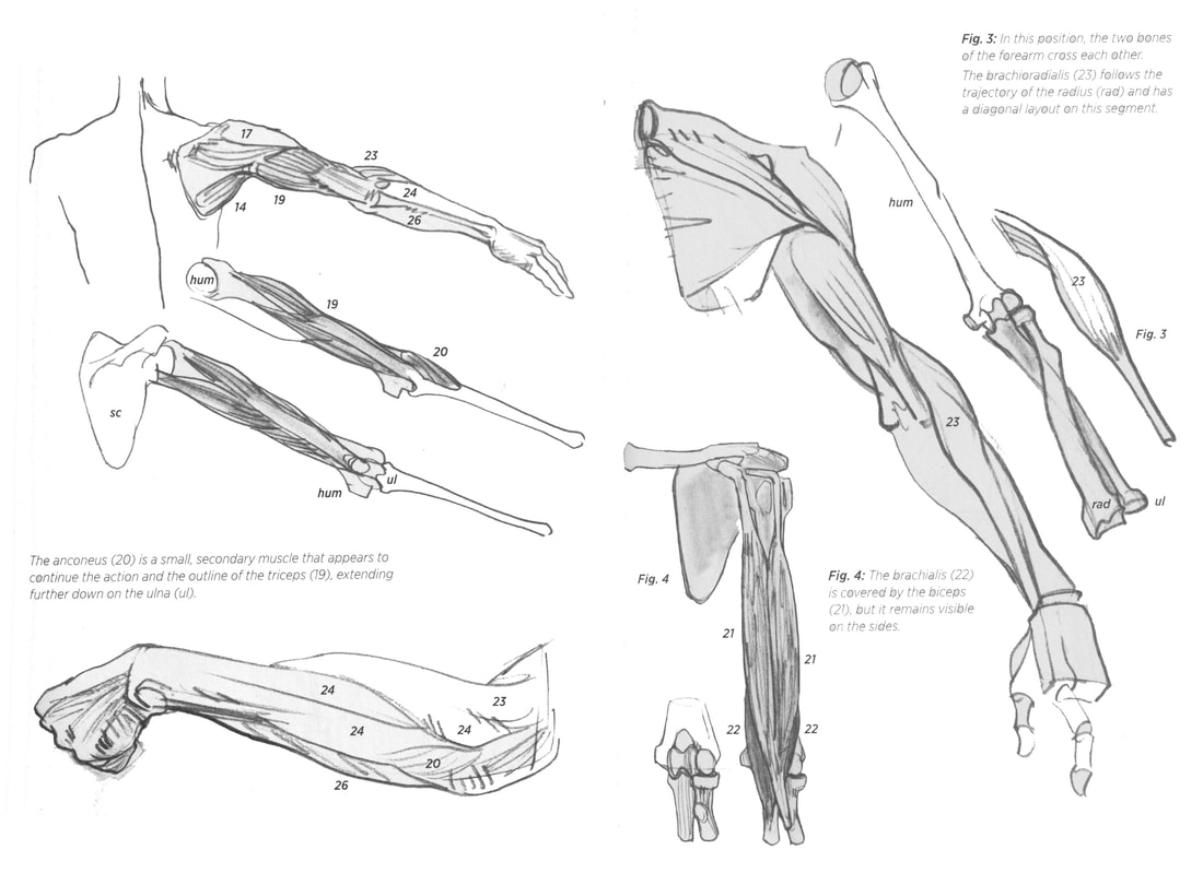
Arms
|
A - Deltoid - ins. scapula, clavicle, humerus - rotating the arm
B - Triceps - ins- scapula, humerus, ulna - extending the forearm C - Brachialis - ins. humerus, ulna - flexing the forearm D - Bicep - ins. scapula, radius - flexing the forearm E - Brachioradialis - ins. humerus, radius - flexing the forearm F - Extensors - extending the hands and fingers |
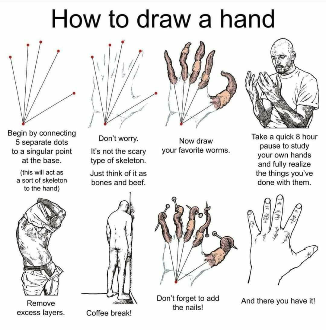
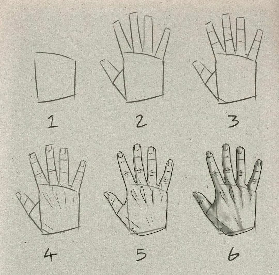
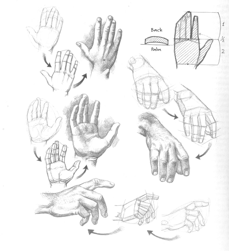
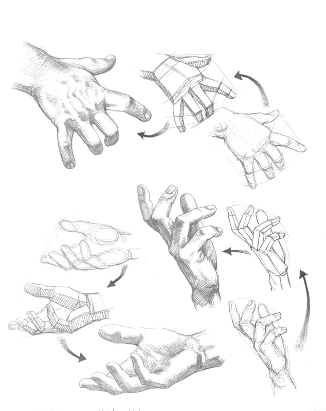
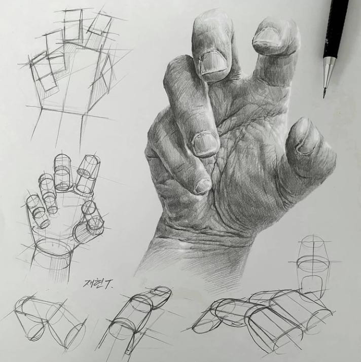
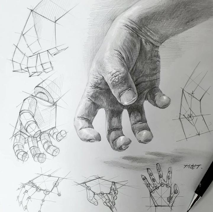
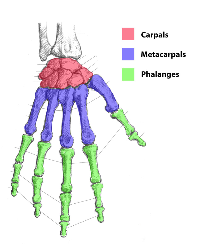
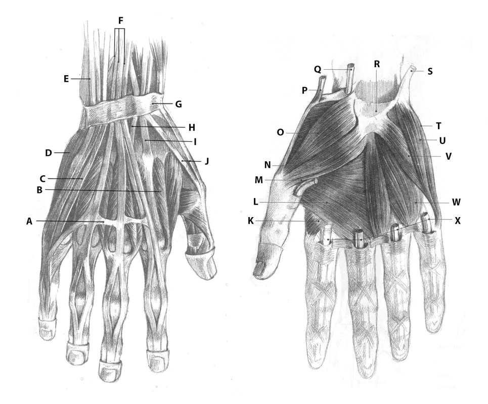
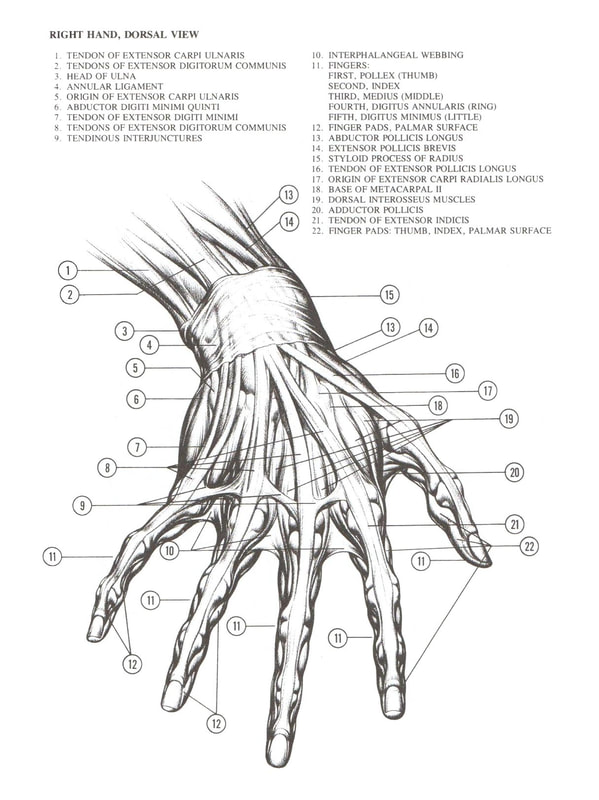
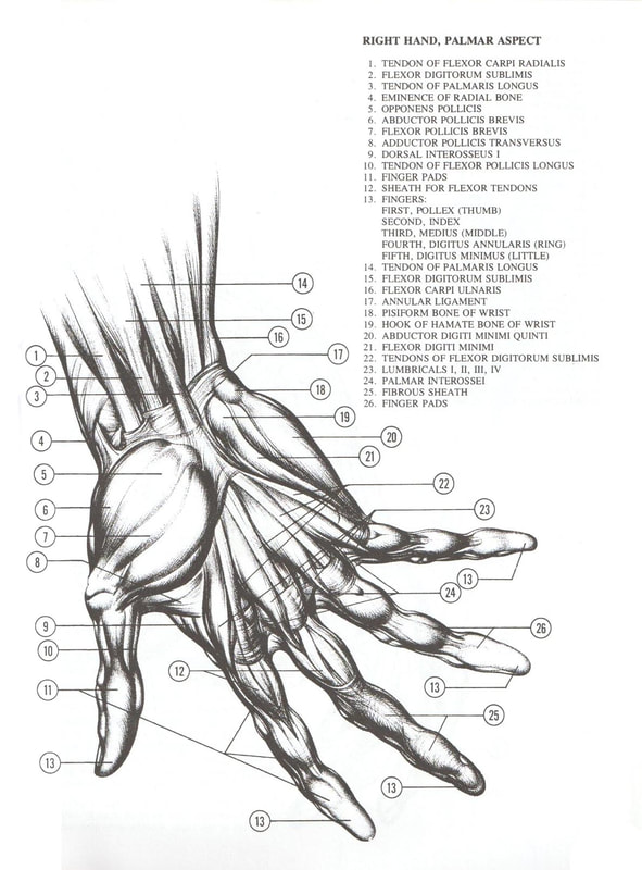
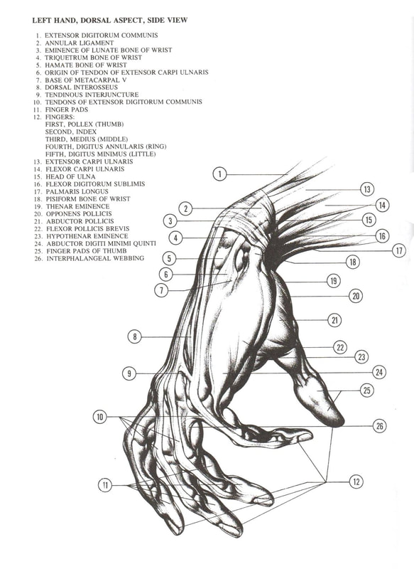
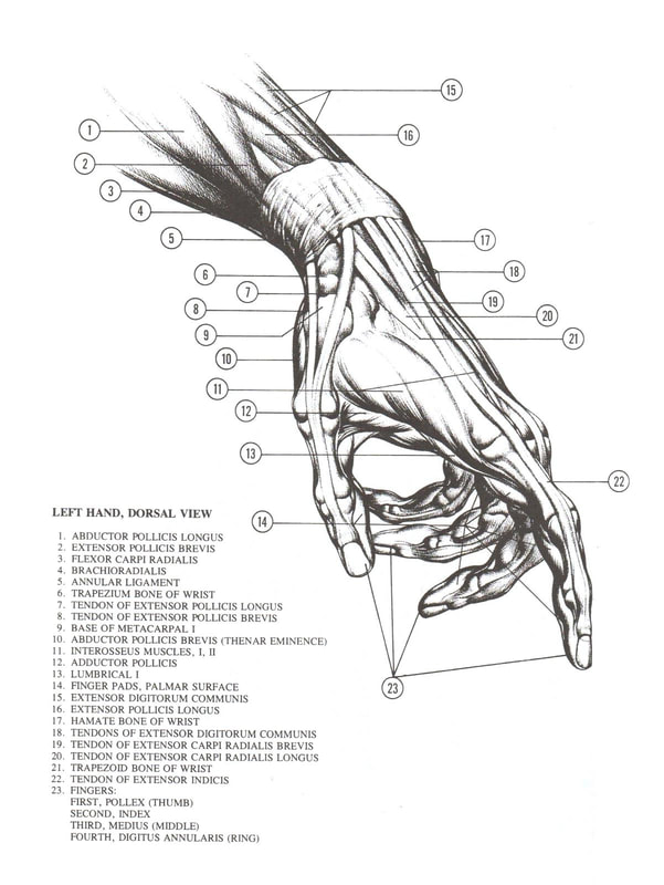
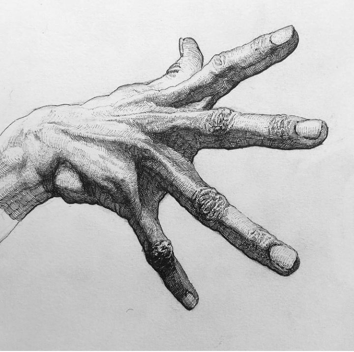
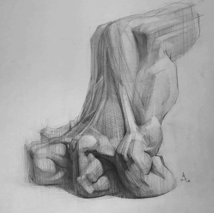
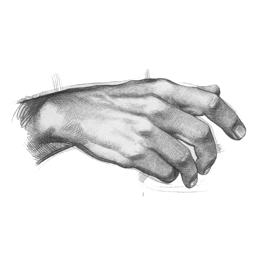
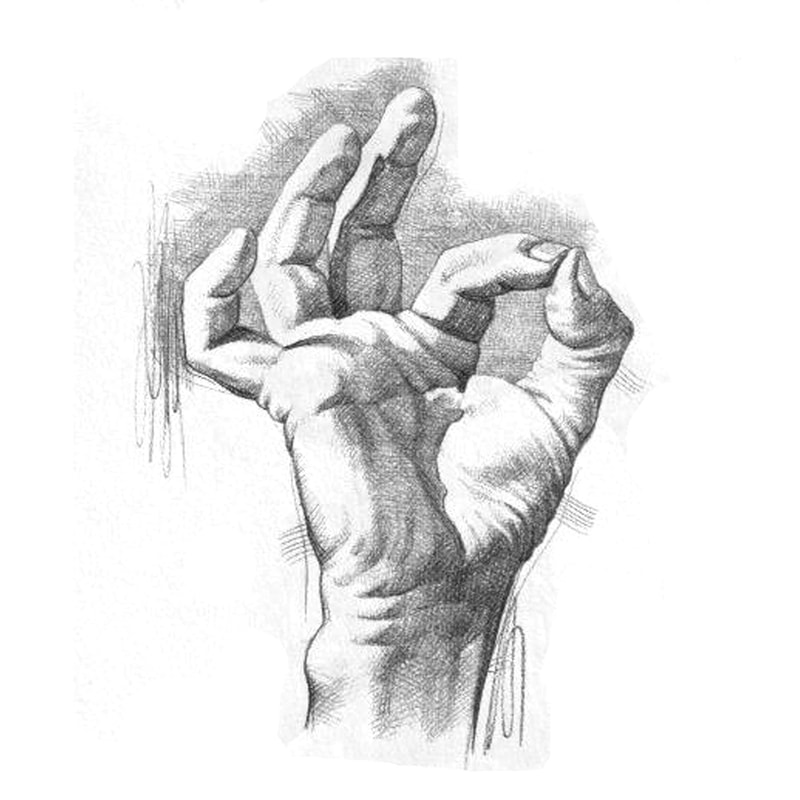
Hands
|
A - Transverse Ligament fastening tendons together
B - Interosseus dorsalis muscle C- Extensor digiti 5th propius muscle D - Abductor digiti 5th muscle E - Extensor carpi ulnaris muscle F - Extensor digitorum communis muscle G - Dorsal retinacular transverse carpal ligament H - Extensor carpi radialis longus muscle |
I - Extensor carpi radialis brevis muscle
J - Extensor digiti 1st longus and brevis muscles K - Interosseus dorsalis 2nd muscle L - Adductor digiti 1st muscle (transverse) M - Adductor digiti 1st muscle (oblique) N - Flexor digiti 1st brevis muscle O - Abductor digiti 1st brevis muscle P - Abductor digiti 1st longus muscle Q - Flexor carpi radialis muscle |
R - Transverse carpal ligament
S - Tendon of the flexor carpi ulnaris muscle T - Abductor digiti 5th muscle U - Flexor digiti 5th muscle V - Opponens digiti 5th muscle W - Lumbricalis muscles X - Tendons of the superficial and deep digital flexors |
The gallery below is composed of different views of teh anatomical structure of the hand from Burne Hogarth's book Drawing Dynamic Hands.

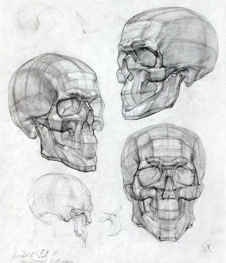
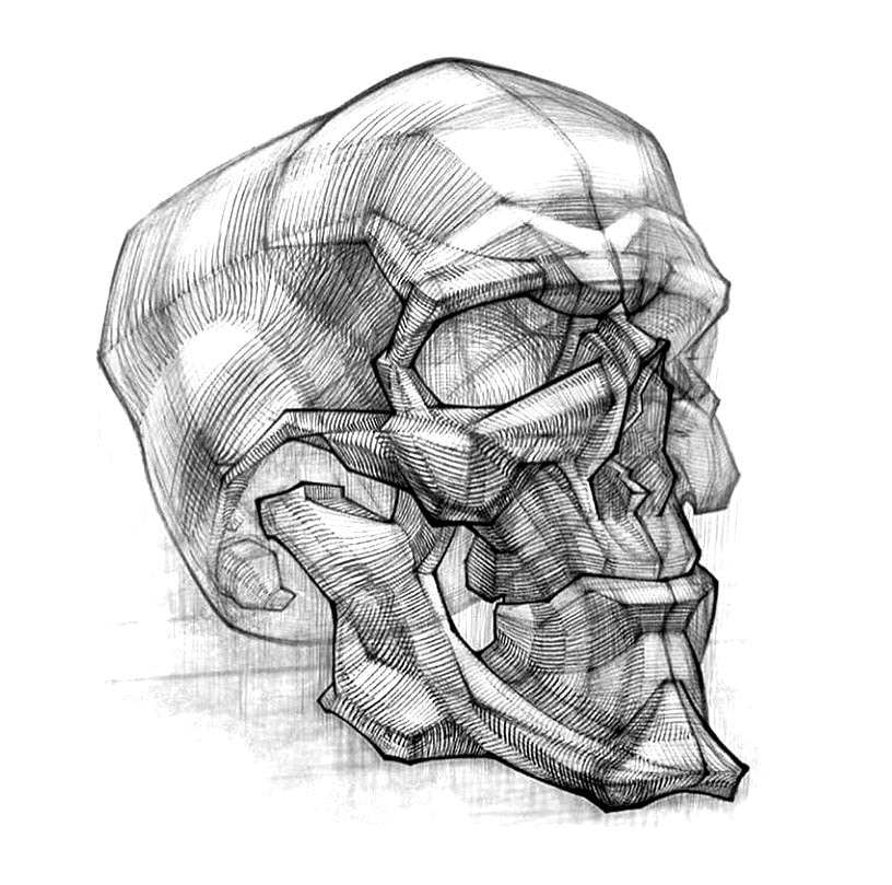
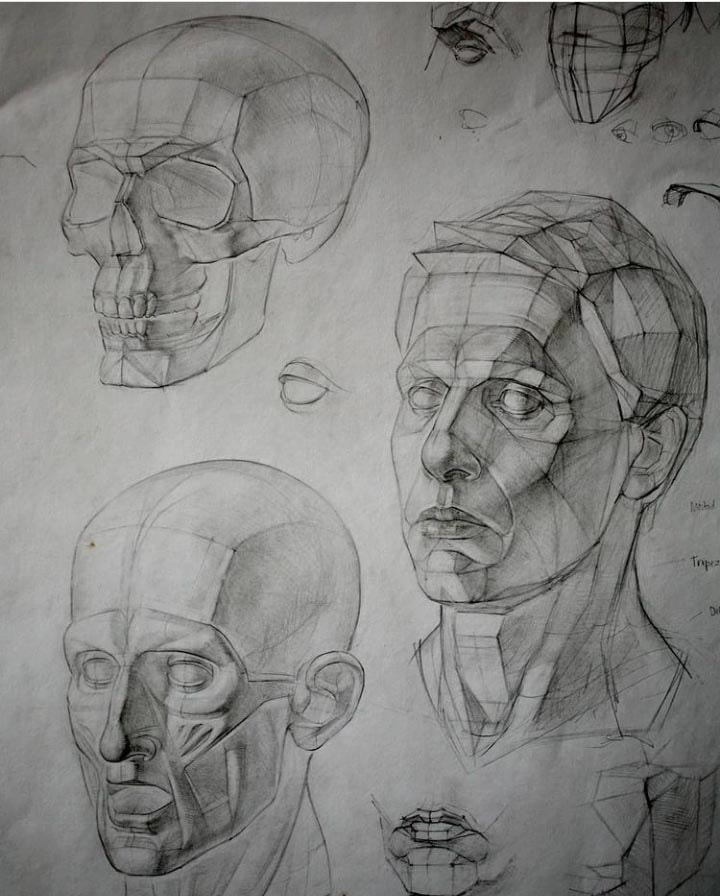
the head
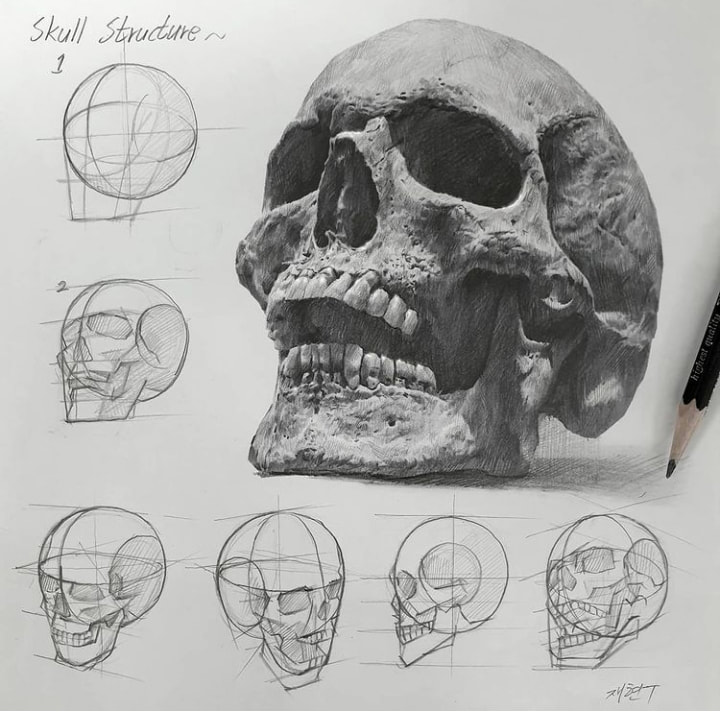
Below are examples of different structural forms from the book Anatomy for Sculptors.
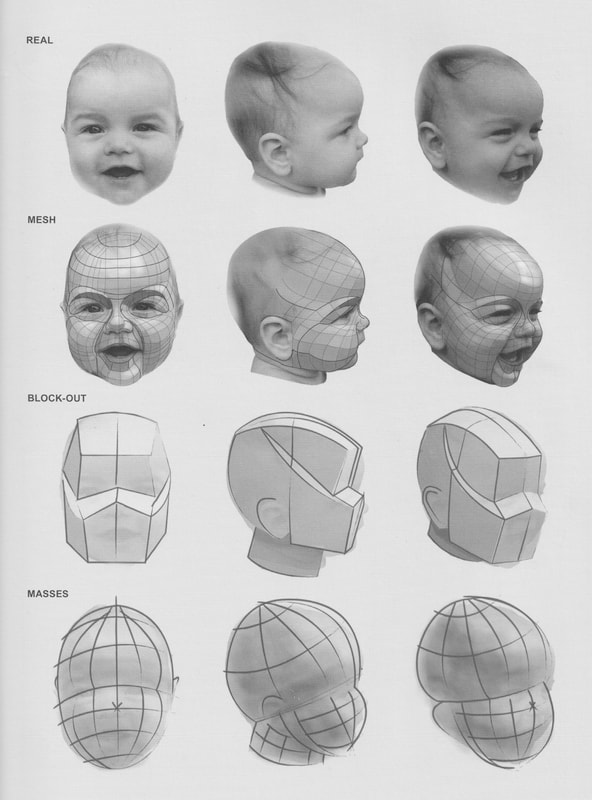
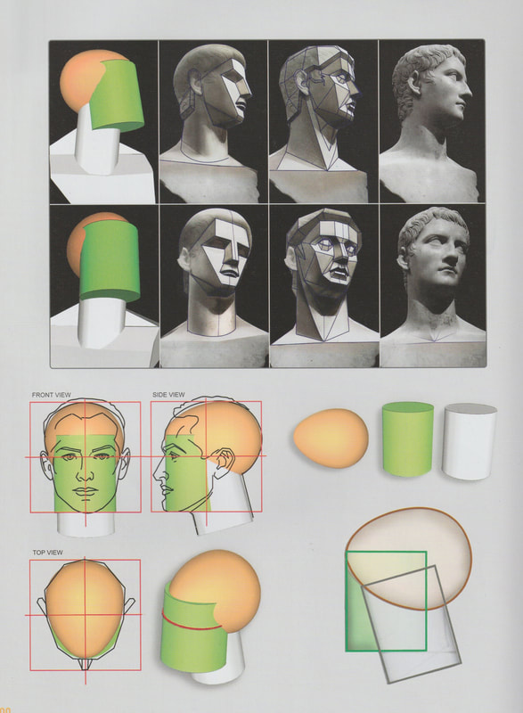
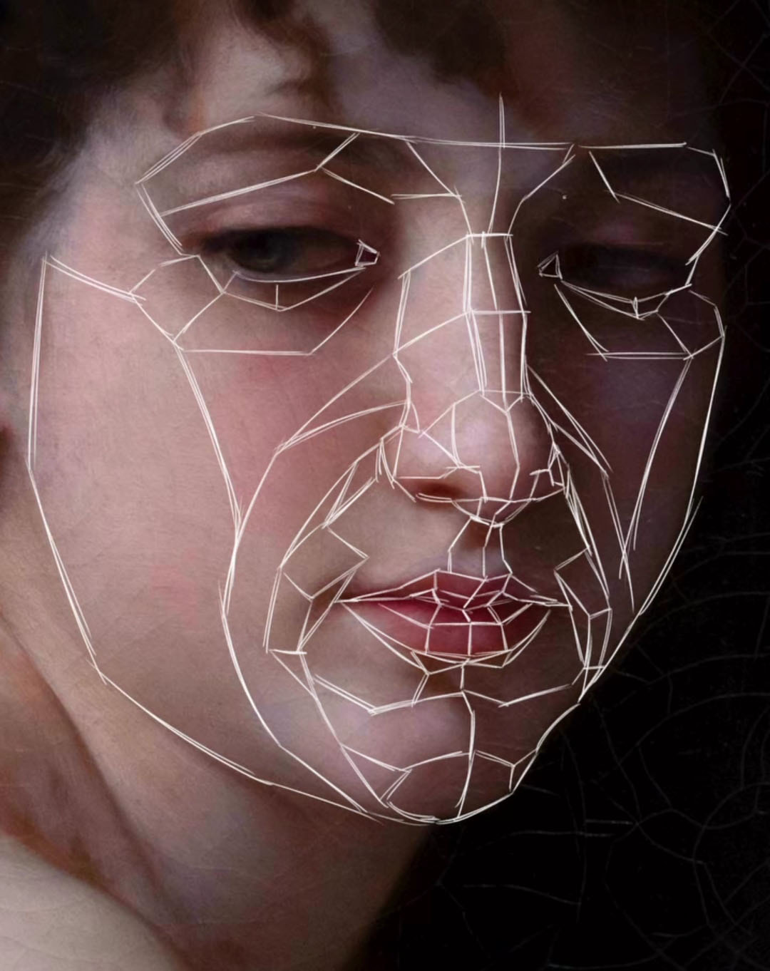
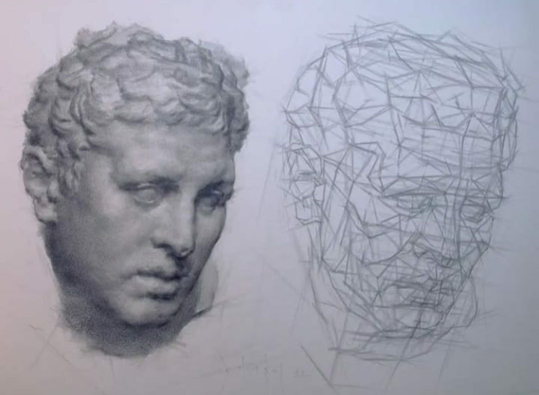
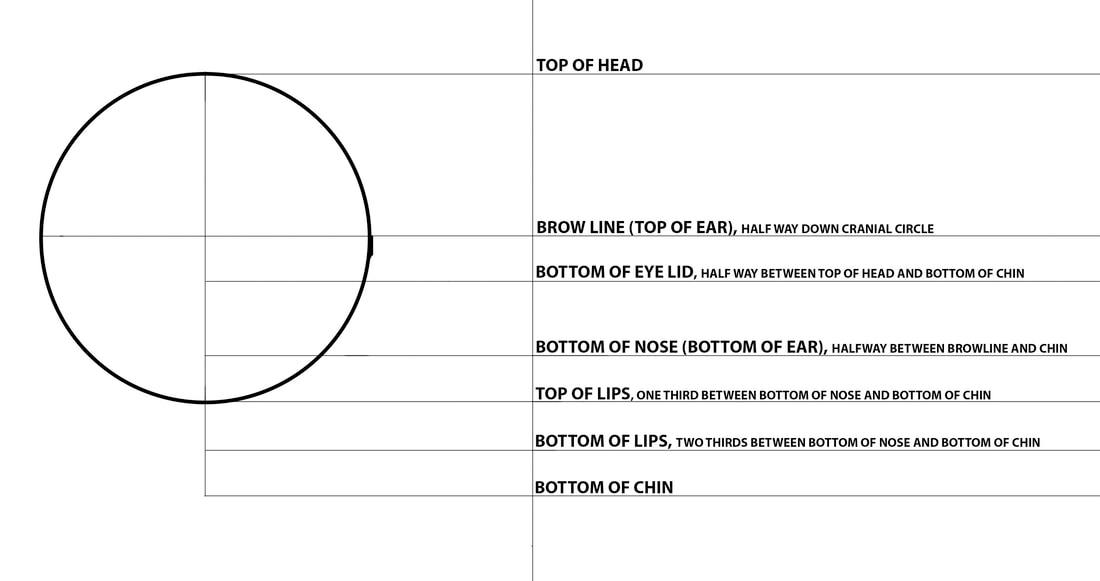
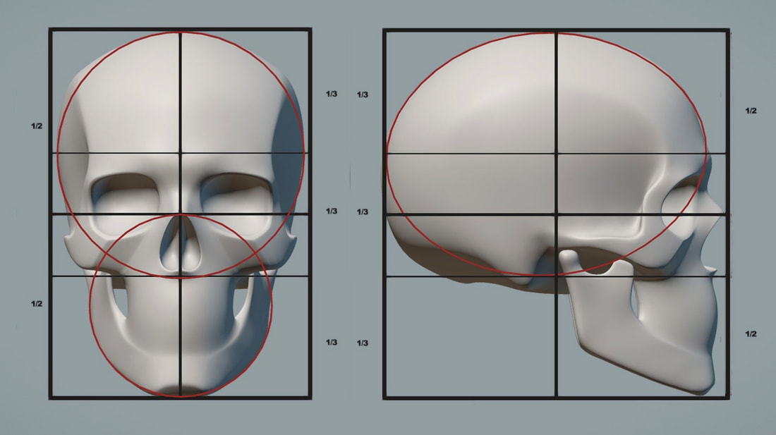
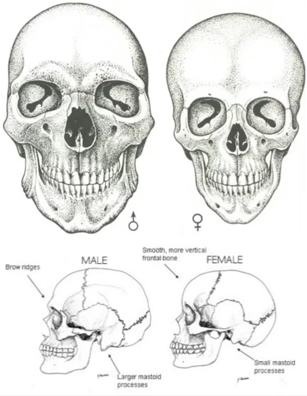
General Proportions of the head
Profile View
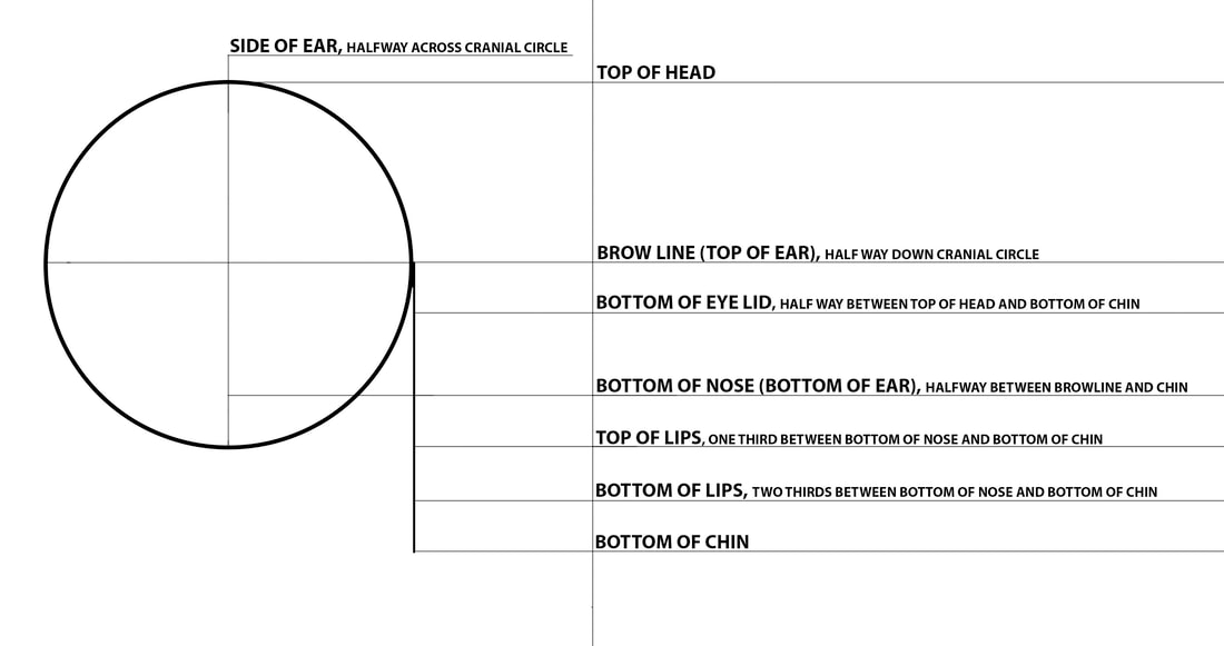
Frontal View
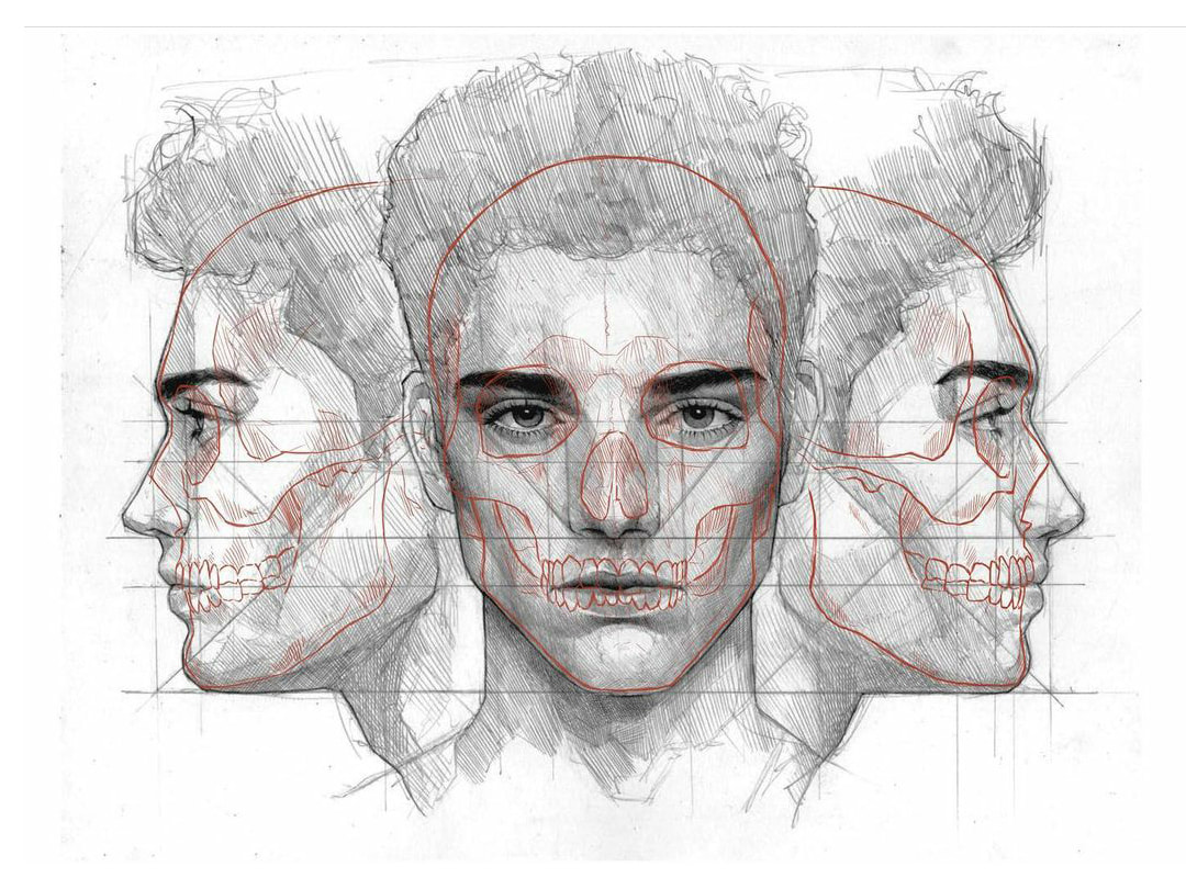
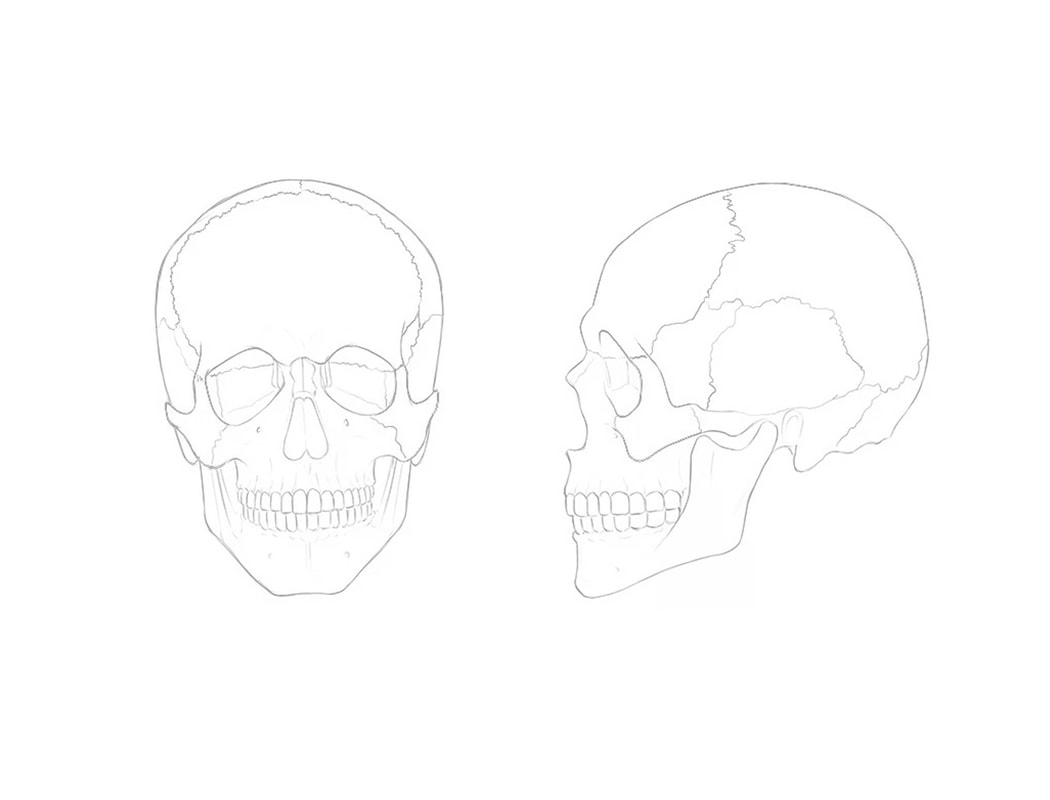
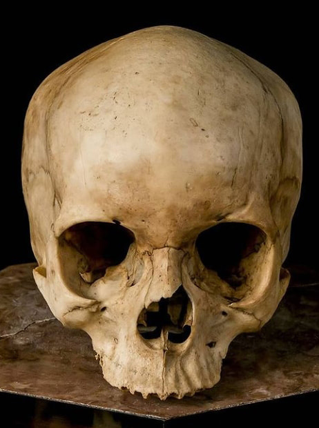
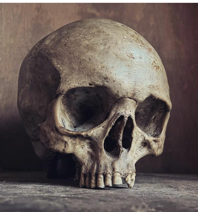
Skull and muscles of the head

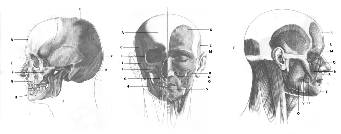
|
A - Frontal
B - Parietal C - Temporal D - Occipital E - Nasal F - Zygomatic G - Maxillary H - Mandible |
I - Mental Protuberance
J - Zygomatic Arch K - Frontalis L - Temporalis M - Orbicularis Oculi N - Levator Labii Superioris Muscles O - Masseter |
P - Occipitalis
Q - Lateralis Nasi R - Zygomaticus S - Orbicularis Oris T - Mentalis U - Buccinator V - Risorius |
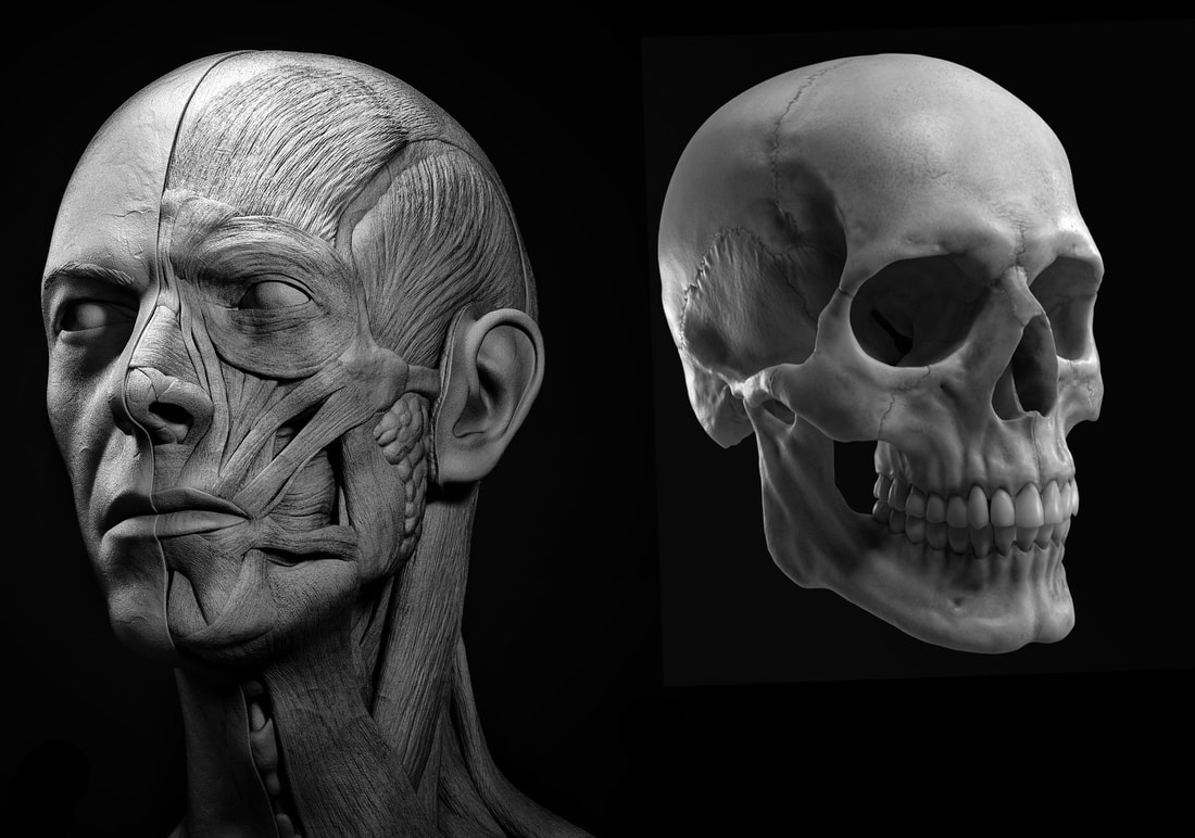
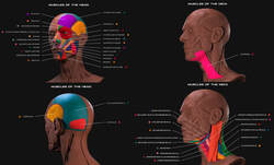
Directly above is a labelled ecorche of the head and neck by Rodrigo Avila and above that is a composite image of an ecorche of the head by Rodrigo Avila next to a human skull for comparison.
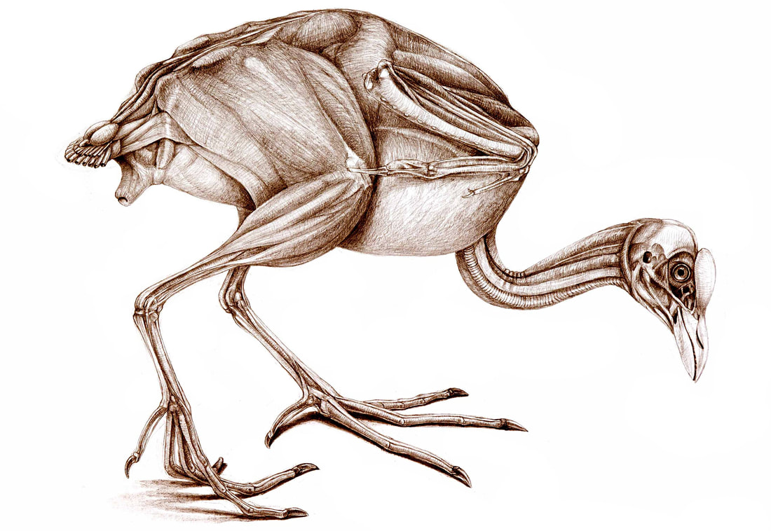
Comparative anatomy
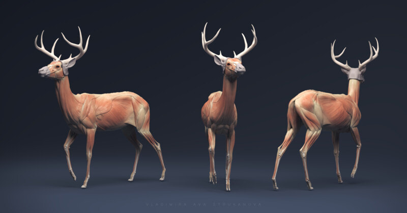
deer
|
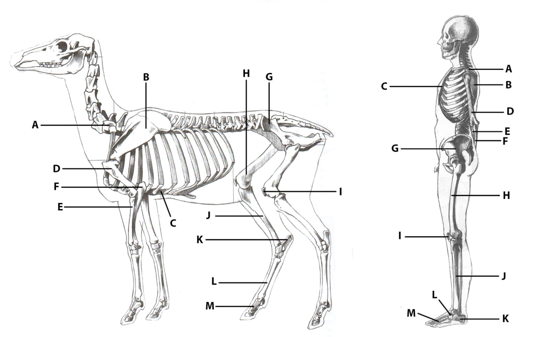
A - C7 Vertebrae
B - Scaplula C - Sternum D - Humerus E - Radius F - Ulna G - Pelvis |
H - Femur
I - Patella J - Tibia K - Calcaneus L - Tarsals M - Phalanges |
Below is a deer ecorche by Vladmira Strukanova.
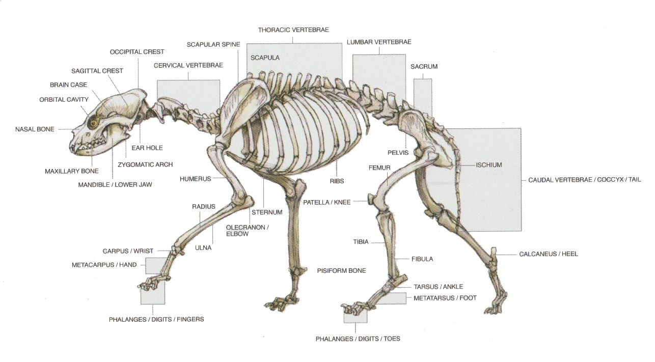
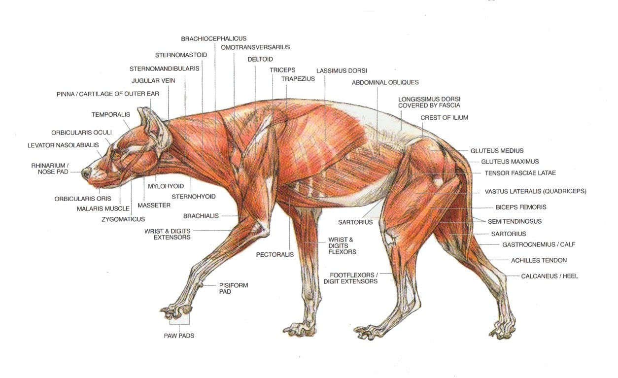
Hyena
The following images of hyena anatomy can be found in in Terryl Whitlatch's book The Science of Creature Design.
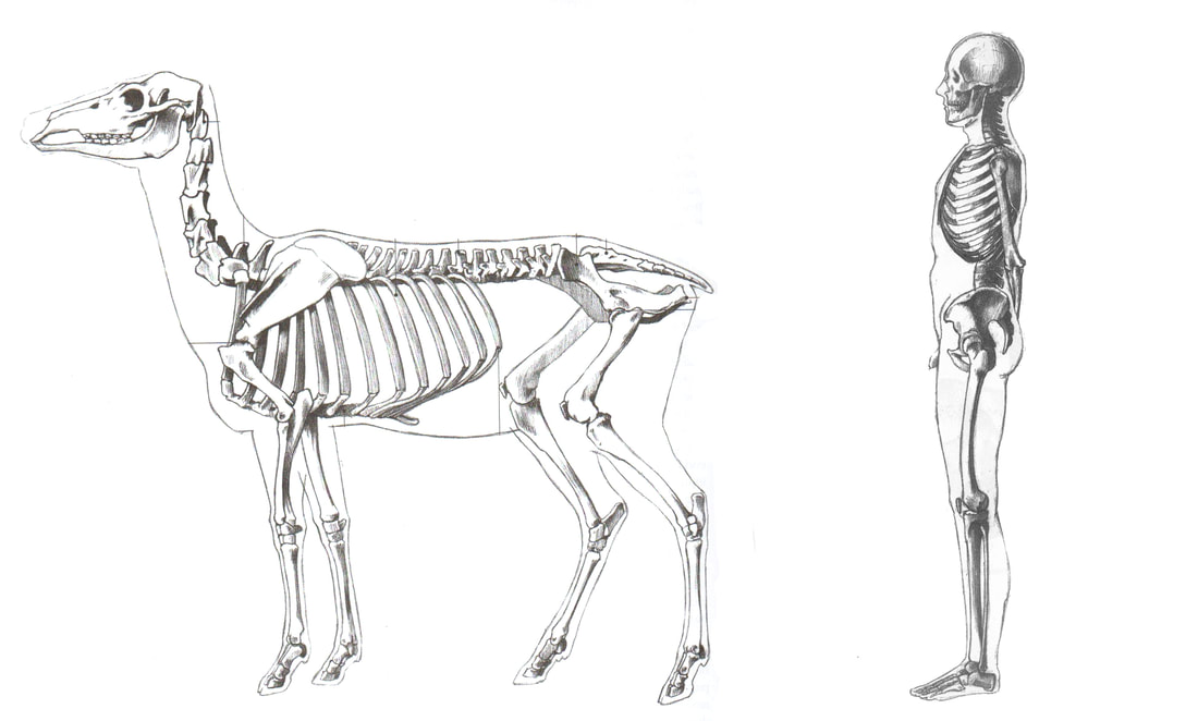
Leg and foot comparisons
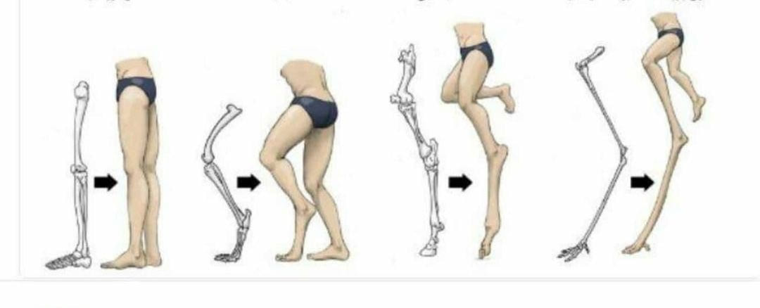
From left to right - Skeletal structure of the human leg, and then as seen with the anatomical structure of a canine, horse, and bird.
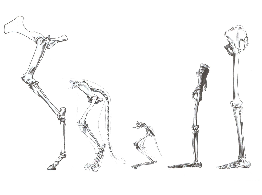
Above are the pelvis and bones of the lower limbs of (from left to right): a horse, lion, house cat, chimpanzee, and human.
Below are the labelled foot bones of a horse, pig, dog, and human.
Below are the labelled foot bones of a horse, pig, dog, and human.
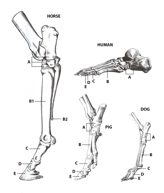
|
A - Tarsal Bones
B - Metatarsal Bones B1 - 3rd metatarsal bone B1 - Splinter bones, 2nd and 4th metatarsal |
C - Proximal phalanx
D - Middle phalanx E - Distal phalanx (nail bone) |
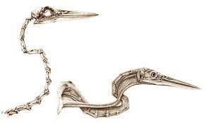
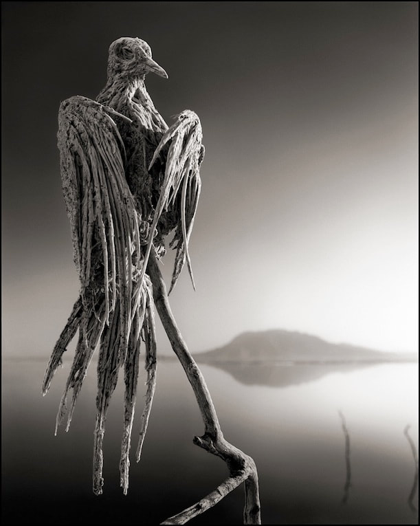
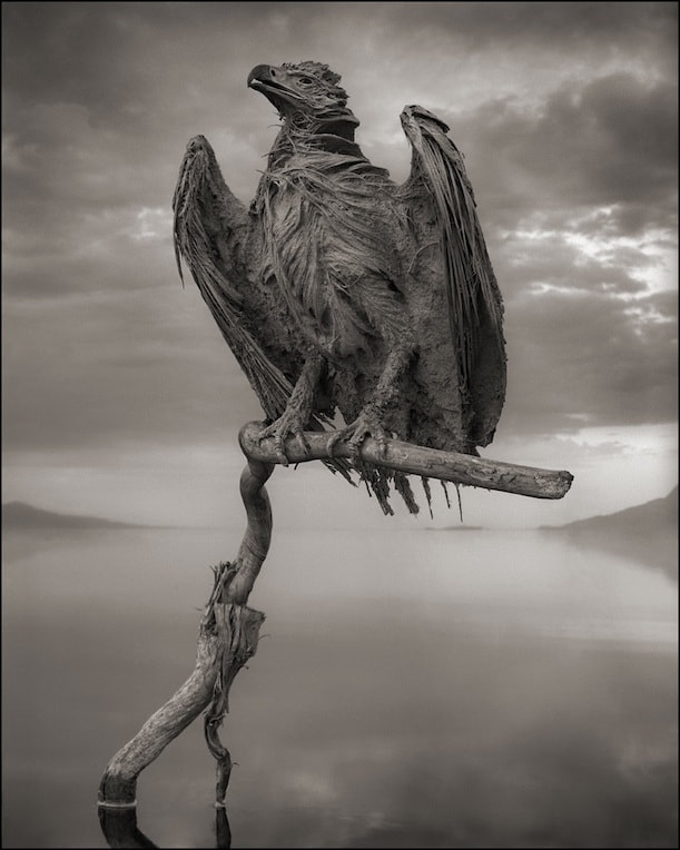
Owls
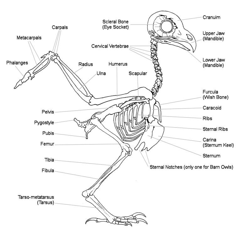
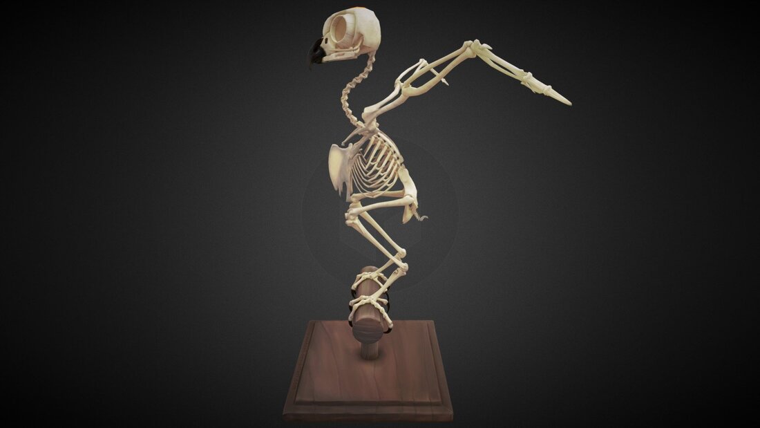

The images below are by Katrina Van Grouw and can be found in their book The Unfeathered Bird.
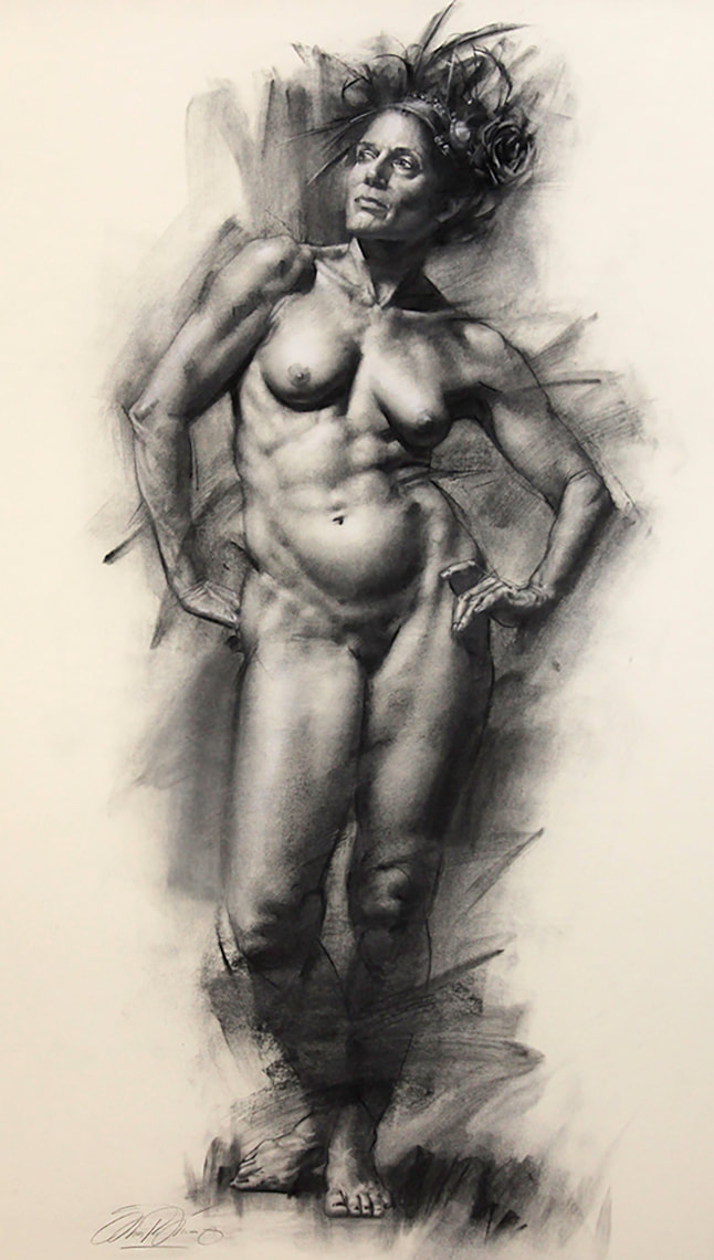
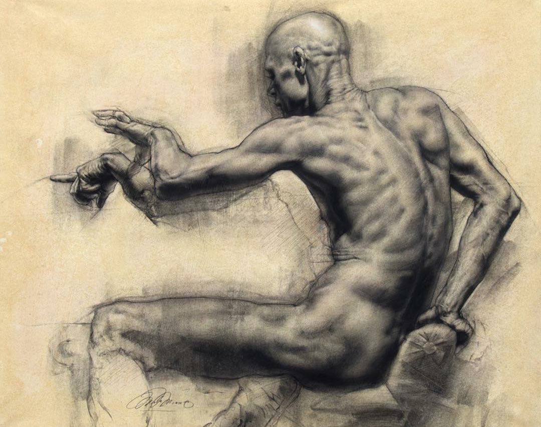
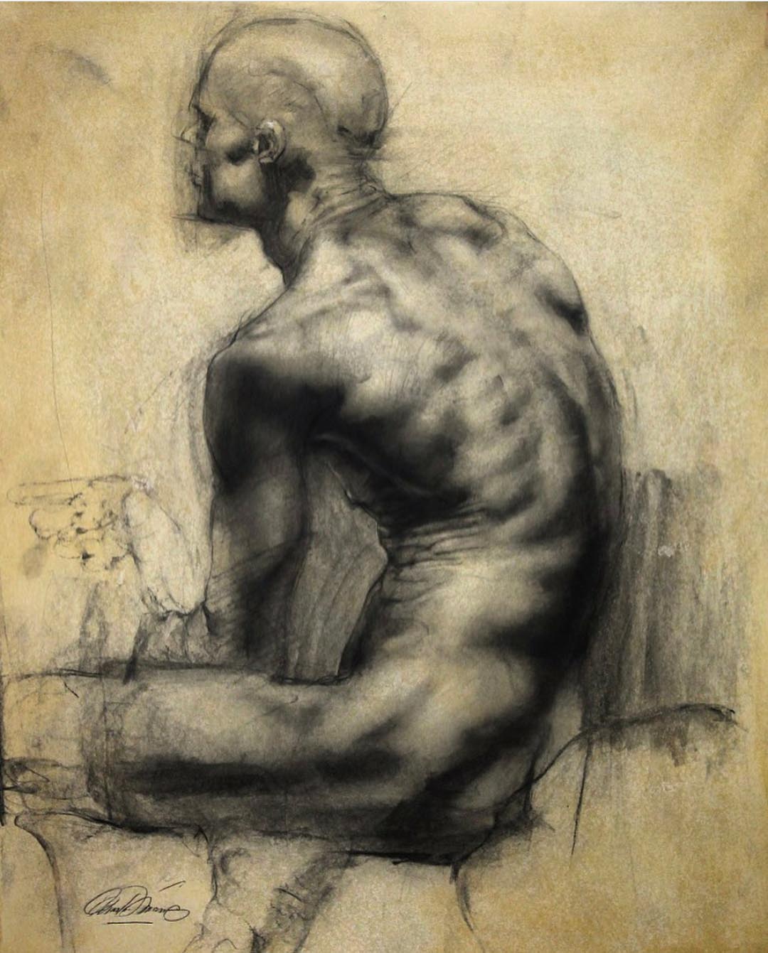
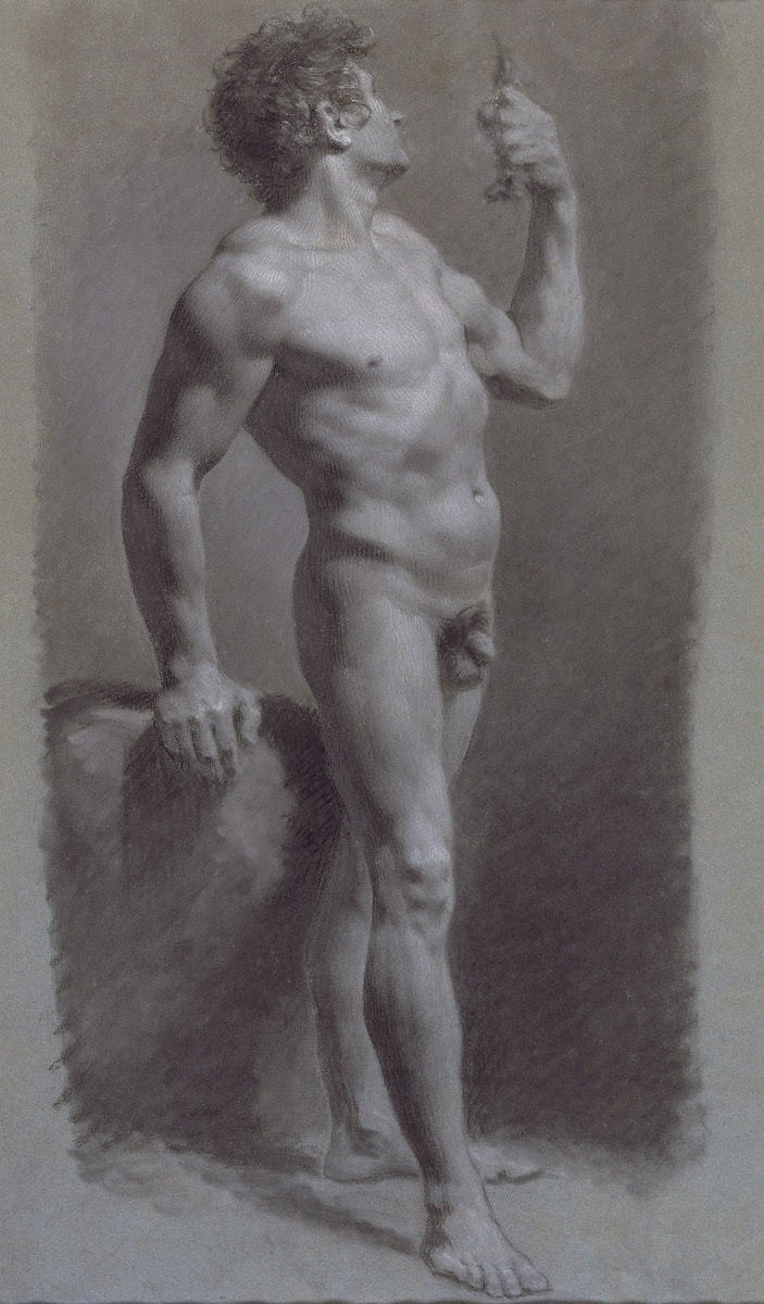
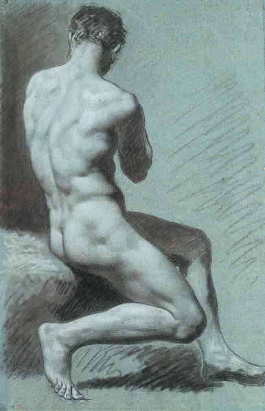
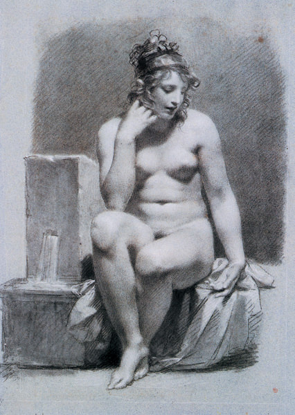
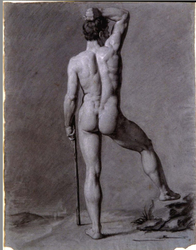
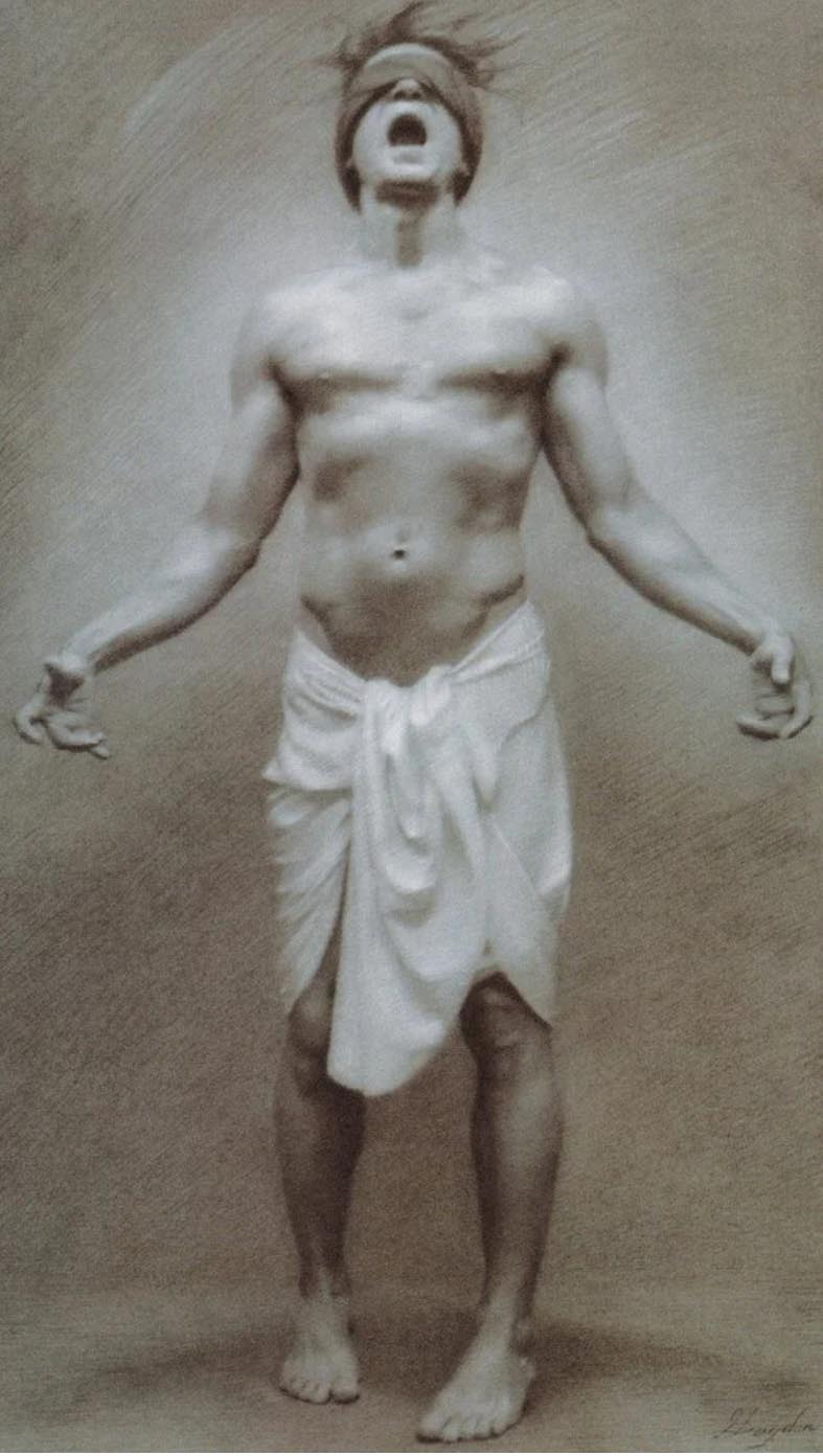
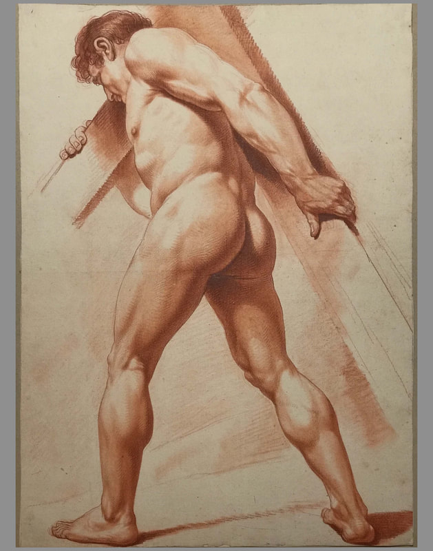
Master Copy Drawings


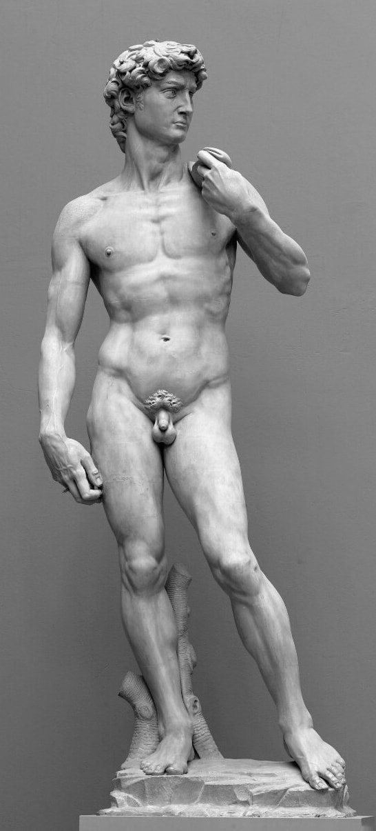
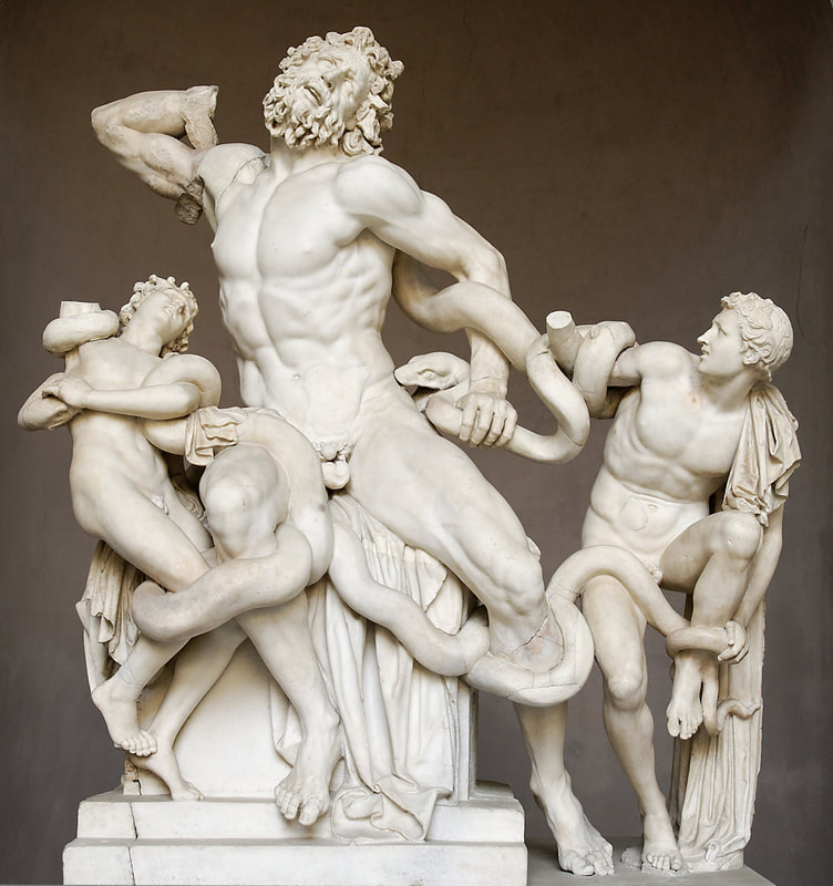
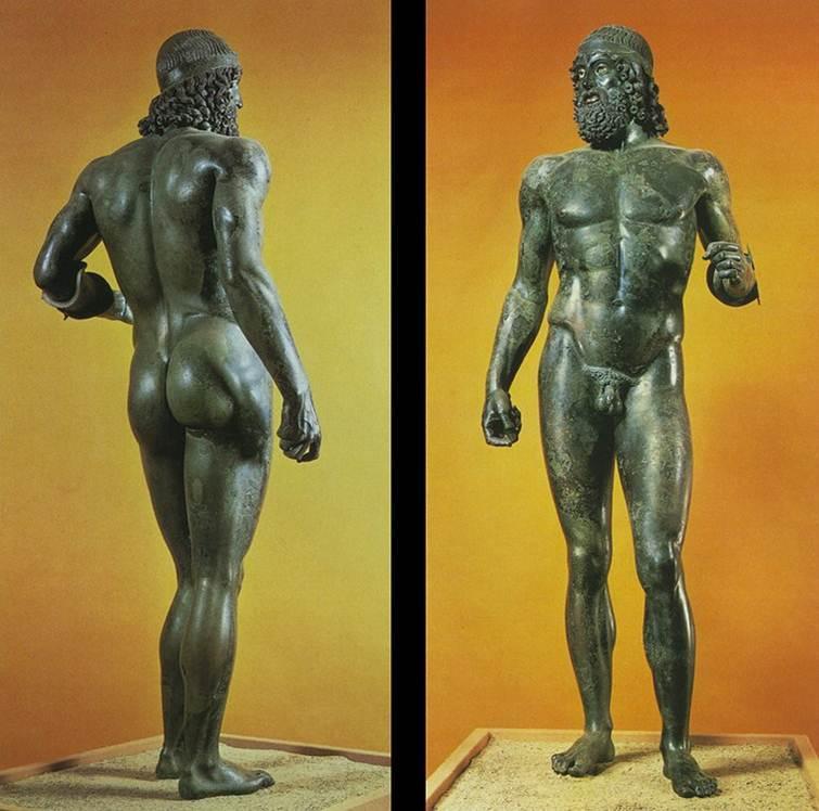
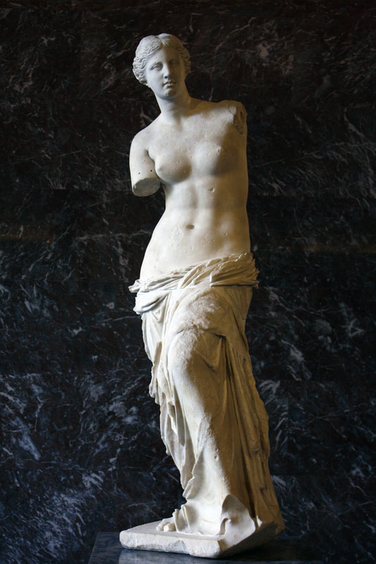
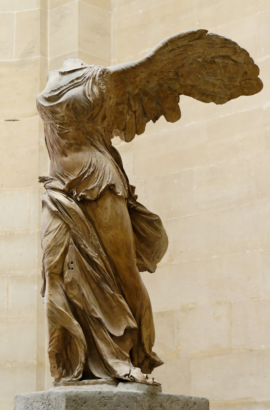
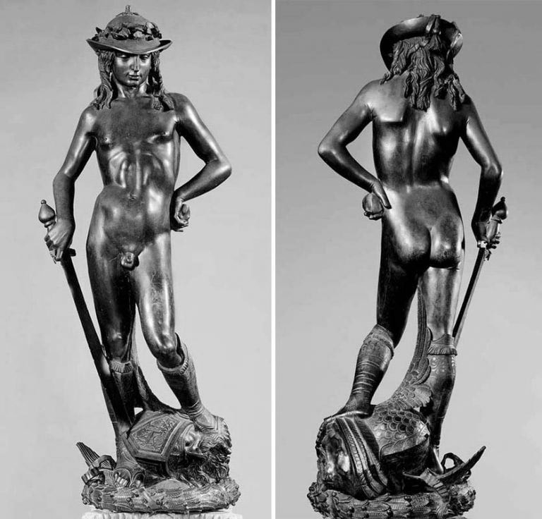
Master Copy Sculptures
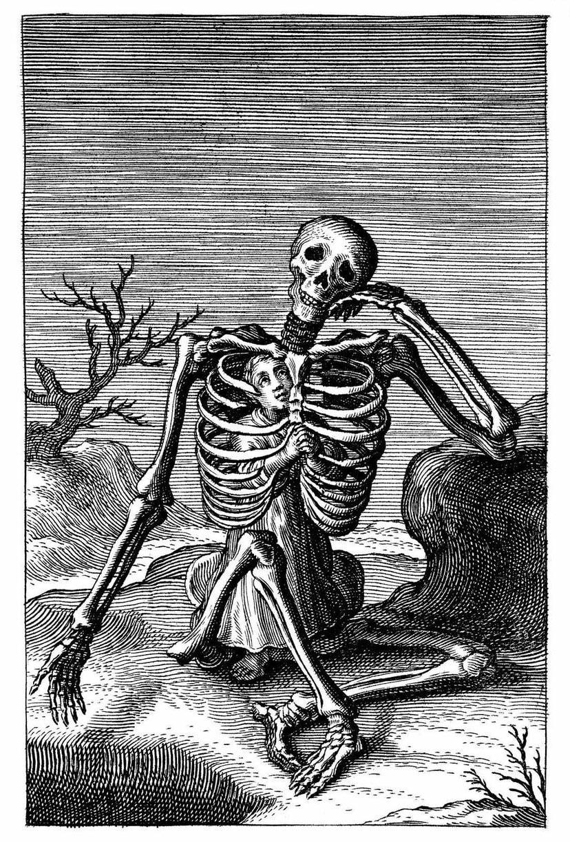
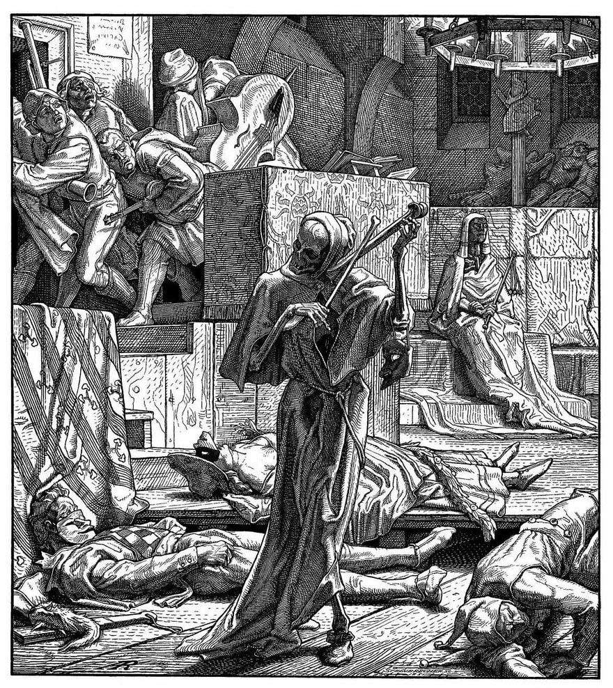
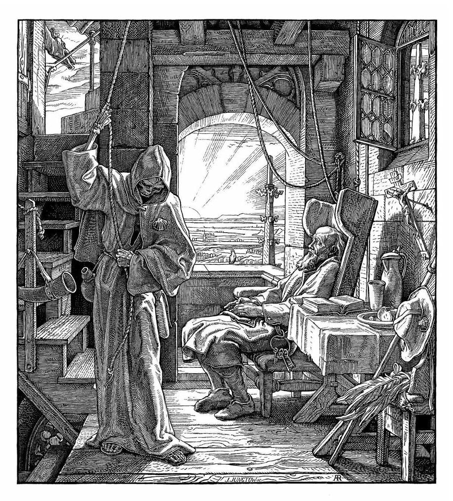
The Ecorche, The Momento Mori, and the Danse Macabre


Skeletons and skulls
Study Sheets


